Copyright
©The Author(s) 2019.
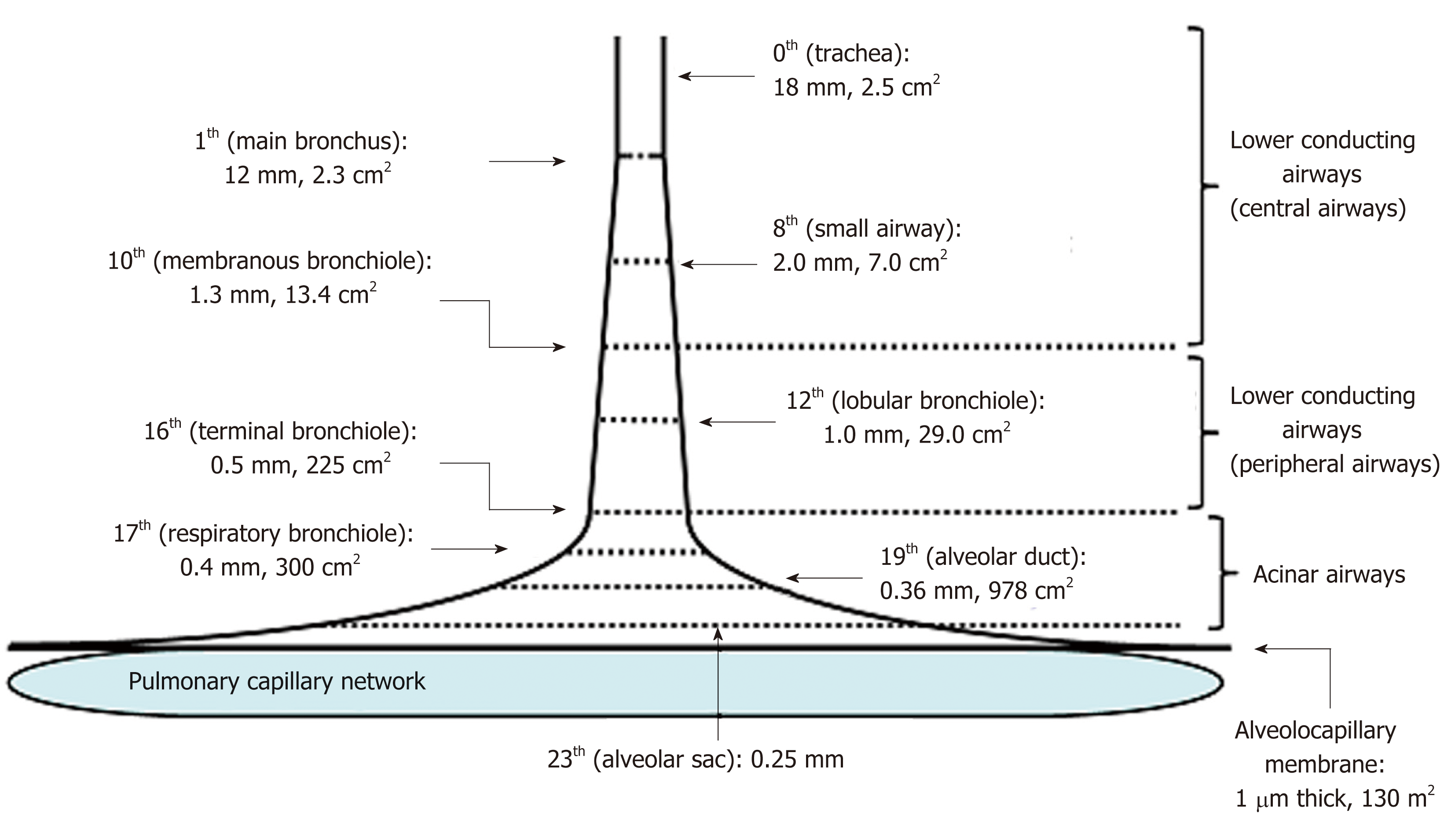
Figure 2 Schematic presentation of effective diameter and cross-sectional area along lower conducting airways and acinar airways (trumpet or thumbtack model).
Generations of branching tree and diameter as well as cross-sectional area in each generation are obtained from model A of Weibel[2]. Lower conducting airways are classified into two routes; i.e., central airways [generation from zero (trachea) to 9th] and peripheral airways (generation from 10th to 16th, in which 16th generation corresponds to terminal bronchiole). Peripheral airways are characterized by membrane bronchioles, whereas small airways are defined by their diameters of less than 2.0 mm originating in the 8th generation. Airways from the 17th generation (respiratory bronchiole) to the 23th generation (alveolar sac) are not included in peripheral airways. Instead, they are defined as acinar (alveolated) airways (acinus indicates that defined by Loeschcke). This is because acinar airways function by carrying gas and exchanging gas. Alveolar sacs end in alveoli formed by alveolar walls containing alveolocapillary membranes and capillary networks. Thickness of the alveolocapillary membrane is about 1.0 μm and total cross-sectional area of the alveolocapillary membrane and capillary network is about 130 m2.
- Citation: Yamaguchi K, Tsuji T, Aoshiba K, Nakamura H, Abe S. Anatomical backgrounds on gas exchange parameters in the lung. World J Respirol 2019; 9(2): 8-29
- URL: https://www.wjgnet.com/2218-6255/full/v9/i2/8.htm
- DOI: https://dx.doi.org/10.5320/wjr.v9.i2.8


