Copyright
©2013 Baishideng Publishing Group Co.
World J Obstet Gynecol. Nov 10, 2013; 2(4): 143-152
Published online Nov 10, 2013. doi: 10.5317/wjog.v2.i4.143
Published online Nov 10, 2013. doi: 10.5317/wjog.v2.i4.143
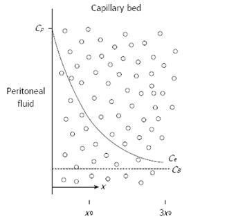
Figure 3 Conceptual diagram of tissue adjacent to the peritoneal cavity.
Solid line shows the exponential decrease in the free tissue interstitial concentration, Ce, as the drug diffuses down the concentration gradient and is removed by loss to the blood perfusing the tissue. Also shown are the characteristic diffusion length, x0, at which the concentration difference between the tissue and the blood has decreased to 37% of its maximum value, and 3x0, at which the difference has decreased to 5% of its maximum value. Cp: The free drug concentration in the peritoneal fluid; CB: The free drug concentration in the blood (or plasma). Modified from Dedrick et al[16].
- Citation: Speeten KVD, Stuart AO, Sugarbaker PH. Pharmacology of cancer chemotherapy drugs for hyperthermic intraperitoneal peroperative chemotherapy in epithelial ovarian cancer. World J Obstet Gynecol 2013; 2(4): 143-152
- URL: https://www.wjgnet.com/2218-6220/full/v2/i4/143.htm
- DOI: https://dx.doi.org/10.5317/wjog.v2.i4.143


