Copyright
©The Author(s) 2017.
World J Orthop. Mar 18, 2017; 8(3): 221-228
Published online Mar 18, 2017. doi: 10.5312/wjo.v8.i3.221
Published online Mar 18, 2017. doi: 10.5312/wjo.v8.i3.221
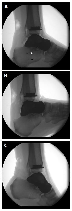
Figure 17 Radiographic examination of the maximum range of motion of the ankle joint after internally bracing of the customized hemiprosthesis.
A: Neutral position; B: Maximum dorsiflexion 22°; C: Maximum plantarflexion 28°. Note the visible bone tunnel in the calcaneus (black arrow) with the interference screw inside the proximal part of the tunnel (white arrow) to prevent tunnel widening by indirect aperture fixation at the subtalar joint performed percutaneously from plantar.
- Citation: Regauer M, Lange M, Soldan K, Peyerl S, Baumbach S, Böcker W, Polzer H. Development of an internally braced prosthesis for total talus replacement. World J Orthop 2017; 8(3): 221-228
- URL: https://www.wjgnet.com/2218-5836/full/v8/i3/221.htm
- DOI: https://dx.doi.org/10.5312/wjo.v8.i3.221


