Copyright
©The Author(s) 2016.
World J Orthop. Oct 18, 2016; 7(10): 638-649
Published online Oct 18, 2016. doi: 10.5312/wjo.v7.i10.638
Published online Oct 18, 2016. doi: 10.5312/wjo.v7.i10.638
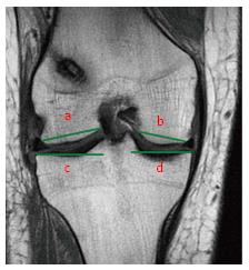
Figure 16 Tibial plateau anatomy is best evaluated by selecting the coronal slice where both tibial spines are visible.
Lateral Femoral Condyle (line a) and Medial Femoral Condyle (line b) diameters are measured from borders of corresponding articular cartilage. Lateral Tibial Plateau (line c) and Medial Tibial Plateau (line d) diameters are measured from the intercondylar spine to the border of the corresponding tibial plateau.
- Citation: Grassi A, Bailey JR, Signorelli C, Carbone G, Wakam AT, Lucidi GA, Zaffagnini S. Magnetic resonance imaging after anterior cruciate ligament reconstruction: A practical guide. World J Orthop 2016; 7(10): 638-649
- URL: https://www.wjgnet.com/2218-5836/full/v7/i10/638.htm
- DOI: https://dx.doi.org/10.5312/wjo.v7.i10.638


