Copyright
©The Author(s) 2016.
World J Orthop. Oct 18, 2016; 7(10): 638-649
Published online Oct 18, 2016. doi: 10.5312/wjo.v7.i10.638
Published online Oct 18, 2016. doi: 10.5312/wjo.v7.i10.638
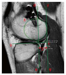
Figure 15 The tibial anterior subluxation with respect to the femur is measured as follow.
With the magnetic resonance imaging acquired in extension and external rotation, the sagittal slice passing through the insertion of the medial gastrocnemius (medial side) or through the most medial cut of the fibula at the tibiofibular joint (lateral side) is selected. Then, a circle over the subchondral line of the posterior condyle (circle a) and a line tangential to the tibial plateau (line b and c) are drawn. The distance (red asterisk) between a perpendicular line to the tibial plateau passing through its posterior margin (line d and e) and a parallel line tangent to the circle (line f and g) is measured.
- Citation: Grassi A, Bailey JR, Signorelli C, Carbone G, Wakam AT, Lucidi GA, Zaffagnini S. Magnetic resonance imaging after anterior cruciate ligament reconstruction: A practical guide. World J Orthop 2016; 7(10): 638-649
- URL: https://www.wjgnet.com/2218-5836/full/v7/i10/638.htm
- DOI: https://dx.doi.org/10.5312/wjo.v7.i10.638


