Copyright
©The Author(s) 2016.
World J Orthop. Oct 18, 2016; 7(10): 638-649
Published online Oct 18, 2016. doi: 10.5312/wjo.v7.i10.638
Published online Oct 18, 2016. doi: 10.5312/wjo.v7.i10.638
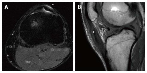
Figure 9 Anterior cruciate ligament reconstruction with bone-patellar tendon bone autograft in a 30-year-old male at 2 years of follow-up.
Donor site pathology is displayed as a hyperintense signal (asterisk) in the proton-density fat saturation axial images surrounding the split patellar tendon (A). In the sagittal proton density weighted slice, the post-operative patellar tendinopathy is displayed as an increase of signal intensity within the tendon itself (asterisk), that resulted in an enlarged and swollen tendon (B).
- Citation: Grassi A, Bailey JR, Signorelli C, Carbone G, Wakam AT, Lucidi GA, Zaffagnini S. Magnetic resonance imaging after anterior cruciate ligament reconstruction: A practical guide. World J Orthop 2016; 7(10): 638-649
- URL: https://www.wjgnet.com/2218-5836/full/v7/i10/638.htm
- DOI: https://dx.doi.org/10.5312/wjo.v7.i10.638


