Copyright
©The Author(s) 2015.
World J Orthop. Aug 18, 2015; 6(7): 505-512
Published online Aug 18, 2015. doi: 10.5312/wjo.v6.i7.505
Published online Aug 18, 2015. doi: 10.5312/wjo.v6.i7.505
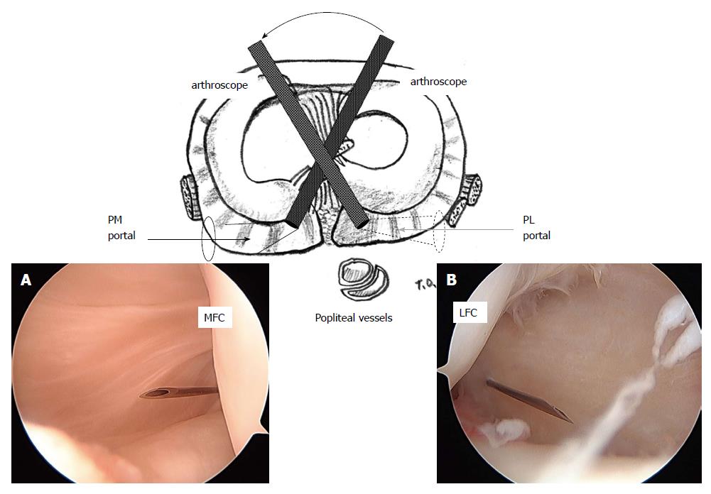
Figure 3 Arthroscopic view of the posteromedial (A) and posterolateral (B) compartment through the intercondylar notch from the anterolateral (A) and anteromedial (B) portals.
A 23-gauge spinal needle is inserted just posterior to the medial (A) and lateral (B) femoral condyle at 5 mm above the tibial articular surface. PM: Posteromedial; PL: Posterolateral; MFC: Medial femoral condyle; LFC: Lateral femoral condyle. (Permission for reproduction was obtained from Nankodo Co., Ltd.).
- Citation: Ohishi T, Takahashi M, Suzuki D, Matsuyama Y. Arthroscopic approach to the posterior compartment of the knee using a posterior transseptal portal. World J Orthop 2015; 6(7): 505-512
- URL: https://www.wjgnet.com/2218-5836/full/v6/i7/505.htm
- DOI: https://dx.doi.org/10.5312/wjo.v6.i7.505


