Copyright
©The Author(s) 2015.
World J Orthop. Aug 18, 2015; 6(7): 505-512
Published online Aug 18, 2015. doi: 10.5312/wjo.v6.i7.505
Published online Aug 18, 2015. doi: 10.5312/wjo.v6.i7.505
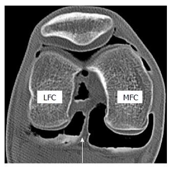
Figure 1 Axial image of double contrast arthrography of the knee.
The white arrow indicates a posterior septum that borders the posterior cruciate ligament anteriorly and posterior capsule posteriorly. Note that posteromedial compartment is larger than posterolateral compartment. LFC: Lateral femoral condyle; MFC: Medial femoral condyle.
- Citation: Ohishi T, Takahashi M, Suzuki D, Matsuyama Y. Arthroscopic approach to the posterior compartment of the knee using a posterior transseptal portal. World J Orthop 2015; 6(7): 505-512
- URL: https://www.wjgnet.com/2218-5836/full/v6/i7/505.htm
- DOI: https://dx.doi.org/10.5312/wjo.v6.i7.505


