Copyright
©2011 Baishideng Publishing Group Co.
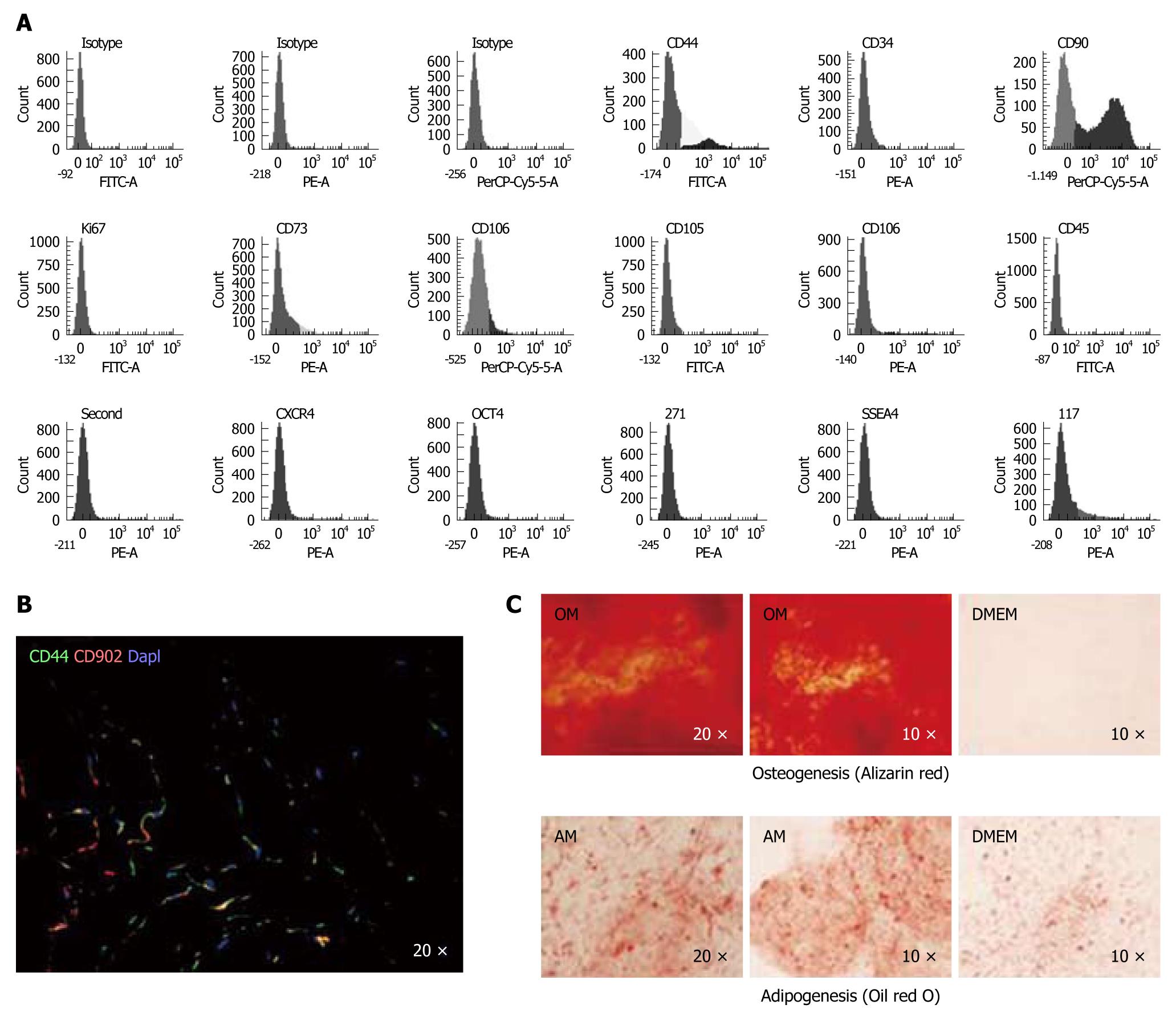
Figure 1 Characterization of the mesenchymal stem cell population from human umbilical cord stroma.
A: Flow cytometry of the principal mesenchymal stem cell (MSC) markers. In each diagram, at the top is the name of the marker, at the bottom the fluorochrome used and at the top right the percentage of positive cells; B: Immunofluorescence analysis of CD44 and CD90 in a human umbilical cord cryosection; DAPI was used to label cell nuclei. (Magnification 20 ×); C: Alizarin red stain (upper) and Oil red O stain (lower) of spheroids engineered from MSCs differentiated in two defined media, proving their pluripotency. OM: Osteogenic medium; AM: Adipogenic medium; DMEM: Control medium with no defined cytokines to promote differentiation.
- Citation: Arufe MC, Fuente ADL, Fuentes I, Toro FJD, Blanco FJ. Umbilical cord as a mesenchymal stem cell source for treating joint pathologies. World J Orthop 2011; 2(6): 43-50
- URL: https://www.wjgnet.com/2218-5836/full/v2/i6/43.htm
- DOI: https://dx.doi.org/10.5312/wjo.v2.i6.43


