Copyright
©The Author(s) 2025.
World J Orthop. Jul 18, 2025; 16(7): 106041
Published online Jul 18, 2025. doi: 10.5312/wjo.v16.i7.106041
Published online Jul 18, 2025. doi: 10.5312/wjo.v16.i7.106041
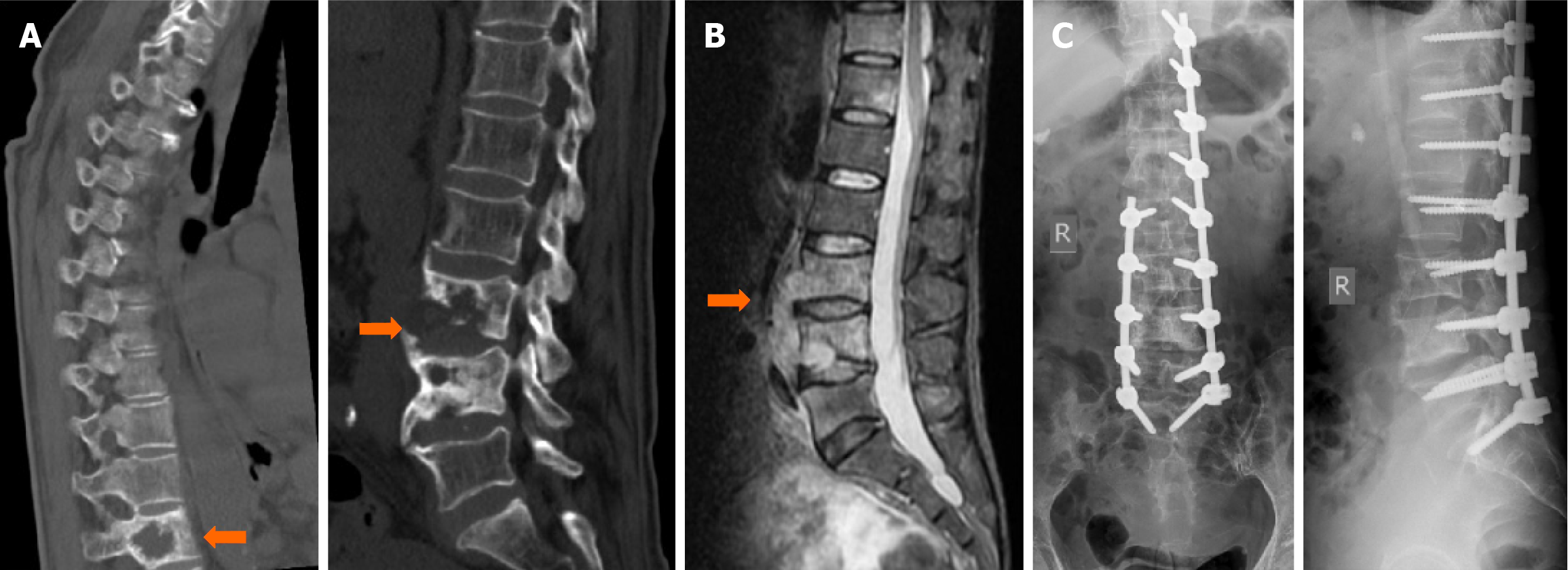
Figure 3 Imaging progression of lumbar tuberculosis treatment.
A: Preoperative computed tomography images showing vertebral bone destruction at T11, L3, and L4; intervertebral space destruction at L3/4; and paravertebral soft tissue swelling; B: Preoperative magnetic resonance imaging showing vertebral bone destruction at L3 and L4 and intervertebral space destruction at L3/4, with associated paravertebral abscess formation; C: Postoperative lumbar X-ray demonstrating intact spinal internal fixation without fracture or displacement, no bone graft migration, and satisfactory intervertebral fusion.
- Citation: Pu FF, Peng XL, Zhou FZ, Zhao XL, Yang L, Cao JQ, Wei L, Feng J, Xia P. Treatment of lumbar tuberculosis with minimally invasive anterior lesion clearance combined with posterior fixation. World J Orthop 2025; 16(7): 106041
- URL: https://www.wjgnet.com/2218-5836/full/v16/i7/106041.htm
- DOI: https://dx.doi.org/10.5312/wjo.v16.i7.106041


