Copyright
©The Author(s) 2025.
World J Orthop. Jun 18, 2025; 16(6): 107753
Published online Jun 18, 2025. doi: 10.5312/wjo.v16.i6.107753
Published online Jun 18, 2025. doi: 10.5312/wjo.v16.i6.107753
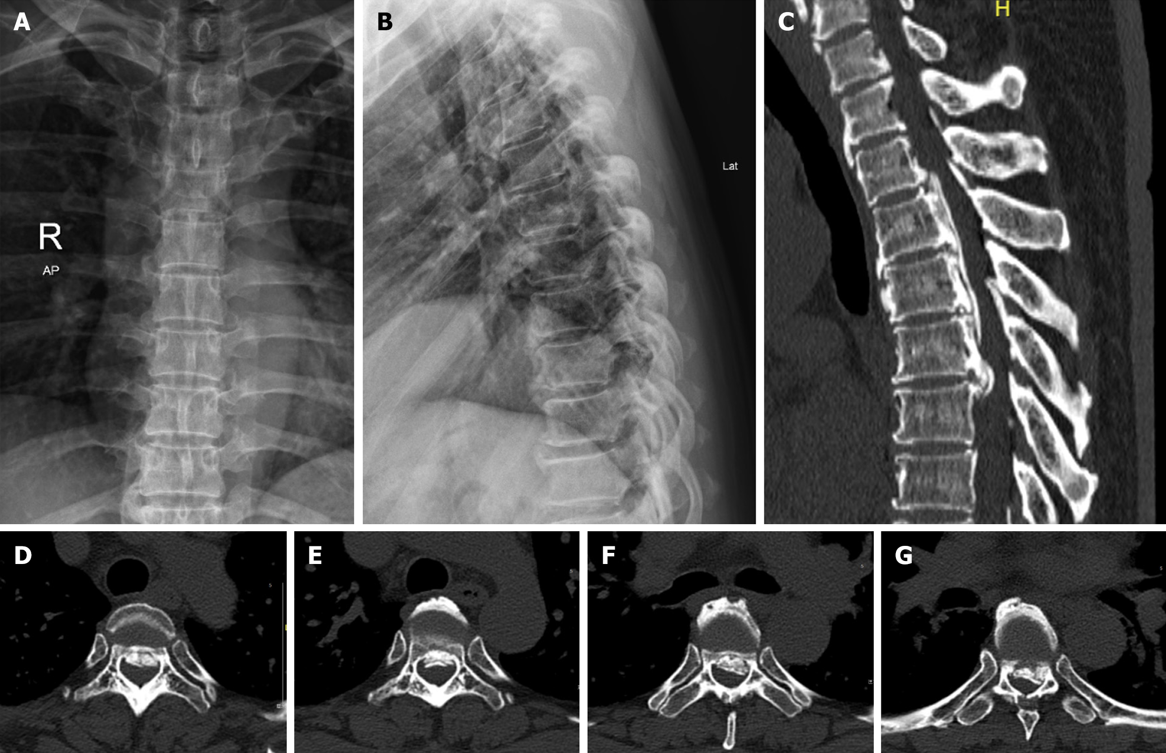
Figure 1 Preoperative diabetic retinopathy and computed tomography.
A and B: Orthoposition and lateral position of thoracic vertebra diabetic retinopathy before operation; C: Thoracic vertebra computed tomography sagittal position before operation. It can be seen that the posterior longitudinal ligament is severely ossified and protrudes backward into the spinal canal; D: Intervertebral space of T2/3; E: Intervertebral space of T3/4; F: Intervertebral space of T4/5; G: Intervertebral space of T5/6.
- Citation: Jin XY, Wang HZ, Yang K, Bao Y, Wang Y, Ben XL, Sun HY. Thoracic anterior controllable antedisplacement fusion for thoracic ossification of the posterior longitudinal ligament: A case report. World J Orthop 2025; 16(6): 107753
- URL: https://www.wjgnet.com/2218-5836/full/v16/i6/107753.htm
- DOI: https://dx.doi.org/10.5312/wjo.v16.i6.107753


