Copyright
©The Author(s) 2025.
World J Orthop. Jun 18, 2025; 16(6): 107397
Published online Jun 18, 2025. doi: 10.5312/wjo.v16.i6.107397
Published online Jun 18, 2025. doi: 10.5312/wjo.v16.i6.107397
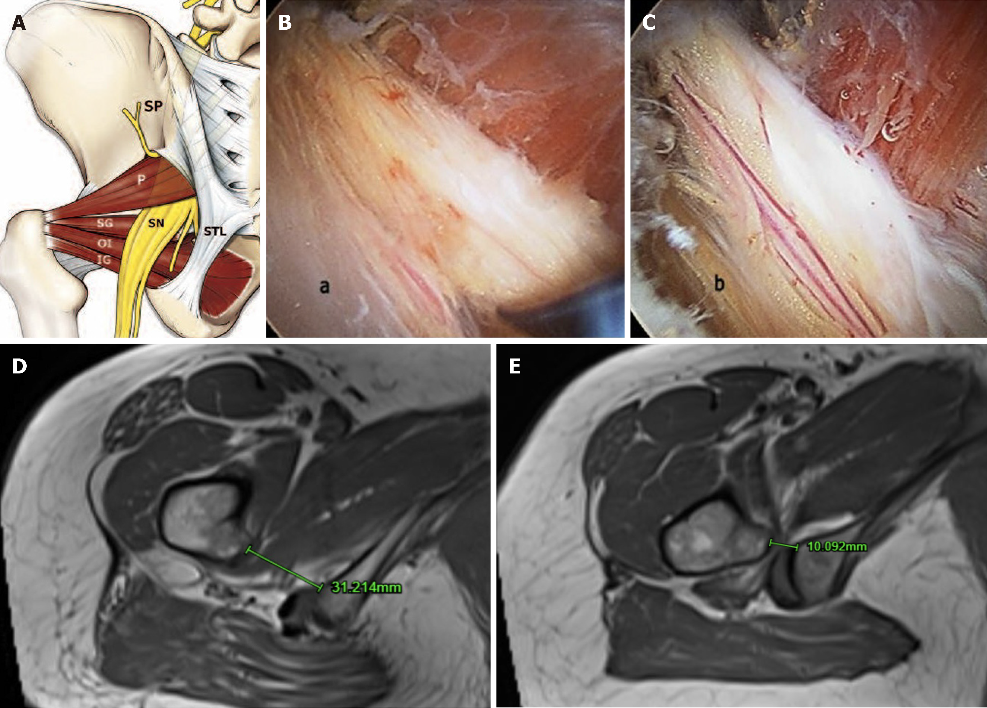
Figure 3 Deep gluteal syndrome and ischiofemoral impingement.
A: Deep gluteal syndrome anatomy; B: Endoscopic view showing edematous and flattened sciatic nerve due to fibrovascular entrapment in a patient with ischemic neuritis; C: Normal vascularization recovery after sciatic nerve neurolysis; D: Normal ischiofemoral space between the medial cortex of the lesser trochanter and lateral cortex of ischial tuberosity (> 20mm); E: Reduced ischiofemoral space signifying underlying ischiofemoral impingement. Figure 3A-C is reproduced from Hernando et al[68]. Copyright © 2015 by Springer Nature. Published by Springer Nature. The authors have obtained the permission for figure using (Supplementary material). SP: Sacral plexus; SN: Sciatic nerve; STL: Sacrotuberous ligament; P: Piriformis muscle; SG: Superior gemellus; OI: Obturator internus muscle; IG: Inferior gemellus muscle.
- Citation: Yeo PY, Kasivishvanaath A, Pérez-Carro L, T J. Top ten causes of non-arthritic hip pain: A comprehensive review. World J Orthop 2025; 16(6): 107397
- URL: https://www.wjgnet.com/2218-5836/full/v16/i6/107397.htm
- DOI: https://dx.doi.org/10.5312/wjo.v16.i6.107397


