
















©The Author(s) 2023.
World J Orthop. Mar 18, 2023; 14(3): 155-165
Published online Mar 18, 2023. doi: 10.5312/wjo.v14.i3.155
Published online Mar 18, 2023. doi: 10.5312/wjo.v14.i3.155
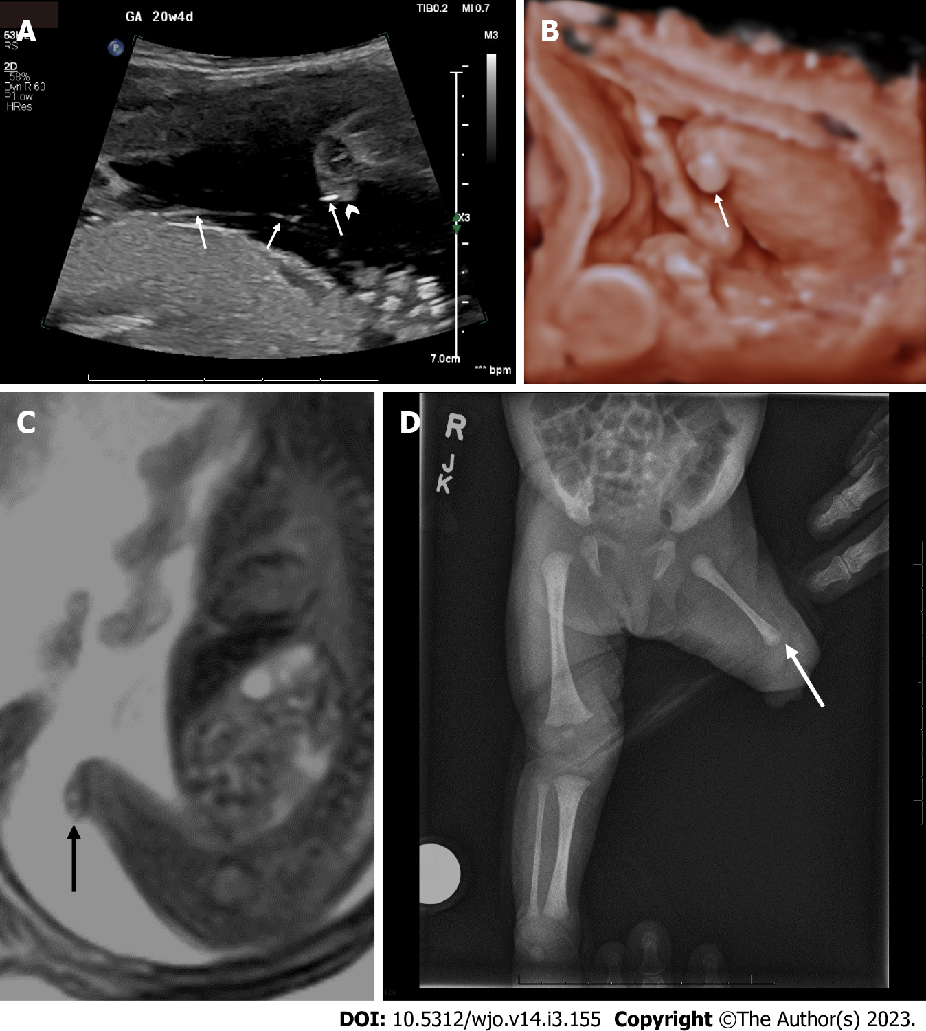
Figure 2 Prenatal images at 20 wk and 4 d and postnatal images demonstrating a congenital transverse limb deficiency below the left knee.
A: Two-dimensional (2D) ultrasound demonstrating the amniotic bands (arrows) that attach to the residual nubbin (arrowhead); B: 3D ultrasound rendered image showing the transverse reduction defect at the level of the knee (arrow); C: Sagittal balanced turbo field echo slice on prenatal MRI of the left lower extremity showing a terminal transverse limb defect (arrow); D: Postnatal AP x-rays of the lower extremities demonstrating the left transverse reduction defect at the left of the knee, residual nubbin (arrow) and shorter left femur compared to right.
- Citation: Vij N, Goncalves LF, Llanes A, Youn S, Belthur MV. Prenatal radiographic evaluation of congenital transverse limb deficiencies: A scoping review. World J Orthop 2023; 14(3): 155-165
- URL: https://www.wjgnet.com/2218-5836/full/v14/i3/155.htm
- DOI: https://dx.doi.org/10.5312/wjo.v14.i3.155