Copyright
©The Author(s) 2023.
World J Orthop. Nov 18, 2023; 14(11): 827-835
Published online Nov 18, 2023. doi: 10.5312/wjo.v14.i11.827
Published online Nov 18, 2023. doi: 10.5312/wjo.v14.i11.827
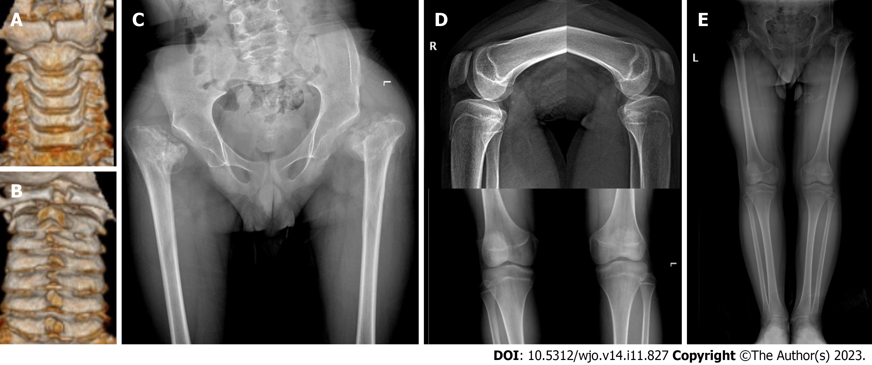
Figure 3 Computed tomography three-dimensional reconstruction of the cervical spine and X-ray of the hip.
A: The view from the front shows dysplasia of the anterior arch of the atlas and the odontoid process of axis; B: The view from the back shows dysplasia of the posterior arch of the atlas; C: A slight right inclination of the pelvis, dysplasia of bilateral hip joints, and severe damage to femoral heads are observed; D: No obvious abnormality is found in the X-ray of bilateral knee joints; E: X-ray of both lower limbs indicate that both lower limbs are equal in length.
- Citation: Jiao Y, Zhao JD, Huang XA, Cai HY, Shen JX. Surgical treatment of atlantoaxial dysplasia and scoliosis in spondyloepiphyseal dysplasia congenita: A case report. World J Orthop 2023; 14(11): 827-835
- URL: https://www.wjgnet.com/2218-5836/full/v14/i11/827.htm
- DOI: https://dx.doi.org/10.5312/wjo.v14.i11.827


