Copyright
©The Author(s) 2021.
World J Orthop. Jun 18, 2021; 12(6): 445-455
Published online Jun 18, 2021. doi: 10.5312/wjo.v12.i6.445
Published online Jun 18, 2021. doi: 10.5312/wjo.v12.i6.445
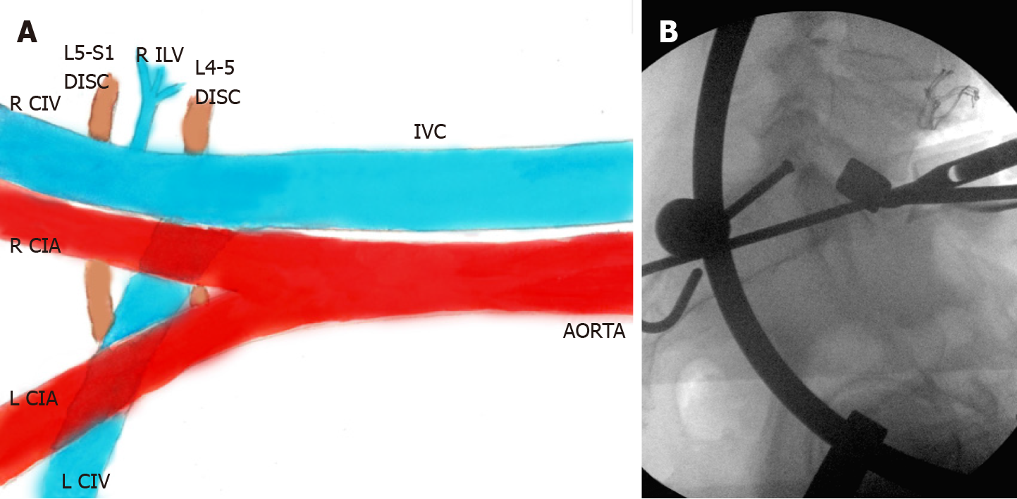
Figure 4 Illustration showing right-sided oblique anterolateral approach (A), and a lateral fluoroscopy image (B) during a right pre-psoas approach.
The right ilio-lumbar vein (ILV) is longer in length and smaller in caliber (A) when compared to the left ILV (Figure 3A). Also note that the right common iliac vein (CIV) is visible throughout in the right pre-psoas approach, unlike the left CIV, which for the most part, is covered by the accompanying left common iliac artery. Image B again identifies specialized trial instrument bent in two planes (similar to Figure 3C and D). IVC: Inferior vena cava; CIV: Common iliac vein; CIA: Common iliac artery; ILV: Ilio-lumbar vein; R: Right; L: Left.
- Citation: Berry CA. Nuances of oblique lumbar interbody fusion at L5-S1: Three case reports. World J Orthop 2021; 12(6): 445-455
- URL: https://www.wjgnet.com/2218-5836/full/v12/i6/445.htm
- DOI: https://dx.doi.org/10.5312/wjo.v12.i6.445


