Copyright
©The Author(s) 2017.
World J Orthop. Nov 18, 2017; 8(11): 846-852
Published online Nov 18, 2017. doi: 10.5312/wjo.v8.i11.846
Published online Nov 18, 2017. doi: 10.5312/wjo.v8.i11.846
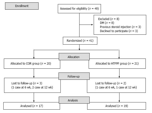
Figure 1 Consort flow diagram showed the randomization process.
COR: Conventional open release; HTFPF: Hypothenar fat pad flap.
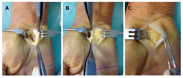
Figure 2 Transverse carpal ligament was exposed.
A: Dissecting the hypothenar fat pad after complete released of transverse carpal ligament (TCL); B: Harvested hypothenar fat pad flap (HTFPF) was prepared to cover the median nerve; C: HTFPF was sutured to radial half of TCL remnant and covered the median nerve.
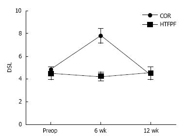
Figure 3 Distal sensory latency was significantly improved in hypothenar fat pad flap group at 6 wk postoperatively, P < 0.
05, but not significant different in between groups at 12 wk postoperative. DSL: Distal sensory latency; COR: Conventional open release; HTFPF: Hypothenar fat pad flap.
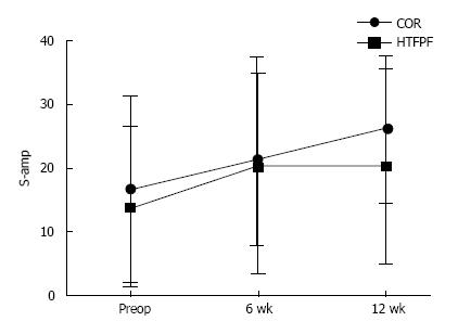
Figure 4 Sensory amplitude was significantly improved in both groups postoperatively.
S-amp: Sensory amplitude; COR: Conventional open release; HTFPF: Hypothenar fat pad flap.
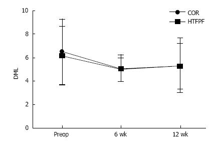
Figure 5 Distal motor latency was not significantly improved postoperatively in both groups and not significant different in between groups.
DML: Distal motor latency; COR: Conventional open release; HTFPF: Hypothenar fat pad flap.
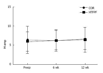
Figure 6 M-amp was not significantly improved postoperatively in both groups and not significant different in between groups.
M-amp: Motor amplitude; COR: Conventional open release; HTFPF: Hypothenar fat pad flap.
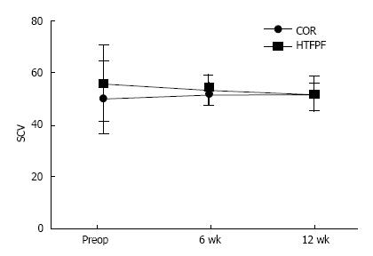
Figure 7 Sensory nerve conduction velocity was not significantly improved postoperatively in both groups and not significant different in between groups.
SCV: Sensory nerve conduction velocity; COR: Conventional open release; HTFPF: Hypothenar fat pad flap.
- Citation: Kanchanathepsak T, Wairojanakul W, Phakdepiboon T, Suppaphol S, Watcharananan I, Tawonsawatruk T. Hypothenar fat pad flap vs conventional open release in primary carpal tunnel syndrome: A randomized controlled trial. World J Orthop 2017; 8(11): 846-852
- URL: https://www.wjgnet.com/2218-5836/full/v8/i11/846.htm
- DOI: https://dx.doi.org/10.5312/wjo.v8.i11.846


