Copyright
©2014 Baishideng Publishing Group Co.
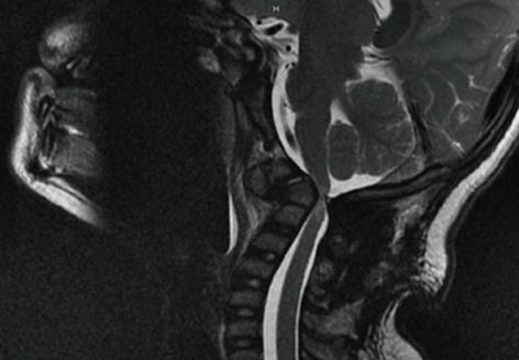
Figure 1 Magnetic resonance imaging showing severe cord compression at the craniocervical junction in a 6 year-old child with instability and myelopathy due to previous failed fusion for atlanto-axial instability.
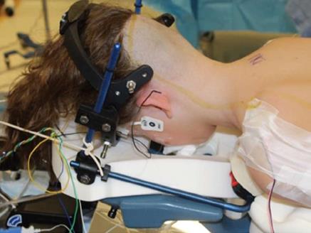
Figure 2 Clinical photo of a 10 year-old child in prone positioning with halo ring and anterior portion of vest attached.
Note the alignment of the head and absence of pressure on the eyes or face.
Figure 3 C1 lateral mass fixation.
A: Axial computed tomography (CT) cut through the atlas. Note the lateral masses and the relationship of the C1 arch meeting the lateral masses which are landmarks for correct starting points for lateral mass screws; B: Sagittal cut of a CT scan in a 14 year-old patient who has underwent C1-C2 arthrodesis with C1-C2 screw rod construct for an odontoid non-union. Note the starting position of the C1 lateral mass screw inferior to the bony arch of C1. The outline of the vertebral artery can be seen on the superior aspect of the C1 arch.
Figure 4 C2 screw fixation.
A: Axial cut computed tomography (CT) scan of intralaminar screw placement at C2. The starting point on the spinous process can be seen as well as the space available for screws; B: Sagittal cut CT of a patient who underwent instrumentation with C2 pars screws. Note the relationship of the vertebral artery in this patient and the need for shorter screw placement; C: Axial CT demonstrating the medial orientation of a C2 pedicle screw with the spinal canal medial and the vertebral artery anterior and lateral.
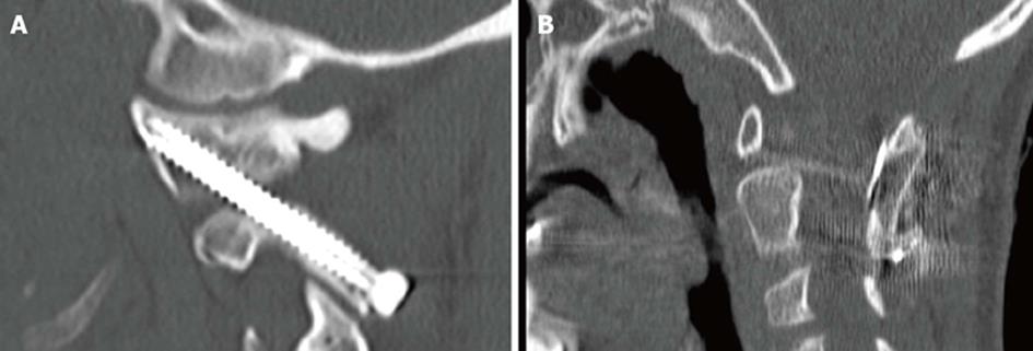
Figure 5 C1-C2 transarticular screws.
A: Sagittal computed tomography (CT) of a fully contained and well placed transarticular screw in a 10 year-old male with Down’s syndrome and os odontoideum; B: Sagittal CT cut demonstrating the placement of a structural iliac crest graft cable grafted in between C1 and C2 which supplemented transarticular screws in an 8 year-old patient with C1-C2 instability.
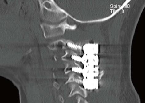
Figure 6 Sagittal computed tomography cut of a 5 year-old patient who underwent lateral mass screw fixation after tumor reconstruction.
- Citation: Hedequist DJ. Modern posterior screw techniques in the pediatric cervical spine. World J Orthop 2014; 5(2): 94-99
- URL: https://www.wjgnet.com/2218-5836/full/v5/i2/94.htm
- DOI: https://dx.doi.org/10.5312/wjo.v5.i2.94


