Copyright
©2014 Baishideng Publishing Group Co.
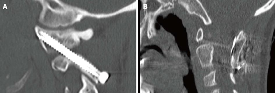
Figure 5 C1-C2 transarticular screws.
A: Sagittal computed tomography (CT) of a fully contained and well placed transarticular screw in a 10 year-old male with Down’s syndrome and os odontoideum; B: Sagittal CT cut demonstrating the placement of a structural iliac crest graft cable grafted in between C1 and C2 which supplemented transarticular screws in an 8 year-old patient with C1-C2 instability.
- Citation: Hedequist DJ. Modern posterior screw techniques in the pediatric cervical spine. World J Orthop 2014; 5(2): 94-99
- URL: https://www.wjgnet.com/2218-5836/full/v5/i2/94.htm
- DOI: https://dx.doi.org/10.5312/wjo.v5.i2.94


