Copyright
©2013 Baishideng Publishing Group Co.
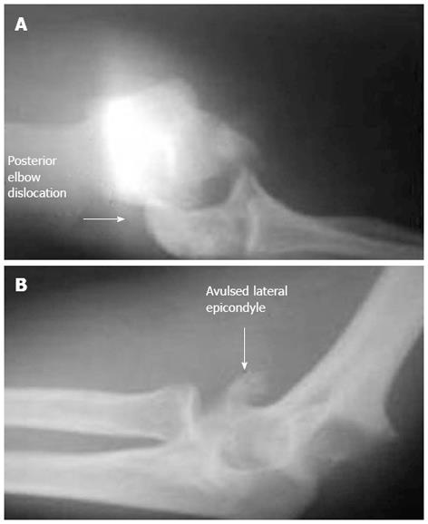
Figure 1 Pre-operative posterior elbow fracture dislocation with avulsed medial and lateral epicondyle fracture radiography.
Figure 2 Intra-operative view.
A: Avulsed medial epicondyle and lateral epicondyle with osteotomized olecranon; B: Avulsed medial epicondyle with nervus ulnaris; C: Plate and screw fixation of olecranon and repaired nervus ulnaris.
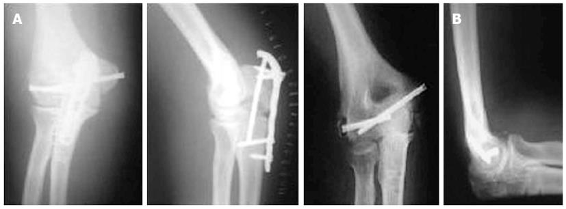
Figure 3 Early post-operative radiography with olecranon plate.
A: Union of the fracture lines, as well as the olecranon osteotomy site, was achieved at the end of forth months post-operatively; B: Elbow radiography after removal olecranon after one year.
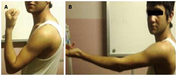
Figure 4 Elbow range of motion after one year.
A: Fully restored flexion; B: Only 20 degrees extension disability.
- Citation: Konya MN, Aslan A, Sofu H, Yıldırım T. Biepicondylar fracture dislocation of the elbow joint concomitant with ulnar nerve injury. World J Orthop 2013; 4(2): 94-97
- URL: https://www.wjgnet.com/2218-5836/full/v4/i2/94.htm
- DOI: https://dx.doi.org/10.5312/wjo.v4.i2.94


