Copyright
©The Author(s) 2023.
World J Orthop. May 18, 2023; 14(5): 362-368
Published online May 18, 2023. doi: 10.5312/wjo.v14.i5.362
Published online May 18, 2023. doi: 10.5312/wjo.v14.i5.362
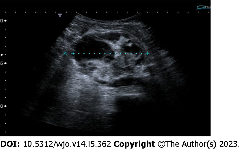
Figure 1 Abdominal ultrasound showed a 7-cm ovoidal lesion with a mixed echo pattern.
It was possible to distinguish fluid and solid components (including diffuse calcifications).
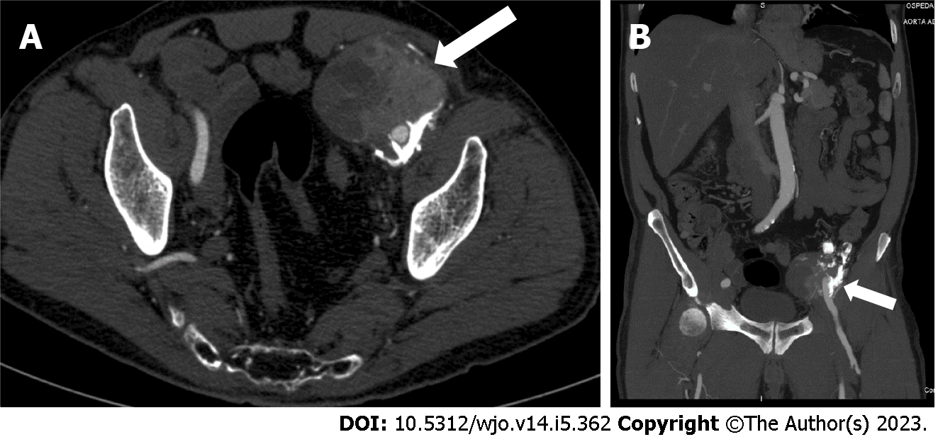
Figure 2 Computed tomography image revealed an 8 cm × 6.
5 cm × 7 cm, highly inhomogeneous mass composed by multiple calcifications at the periphery. A: Axial view; B: Sagittal view.
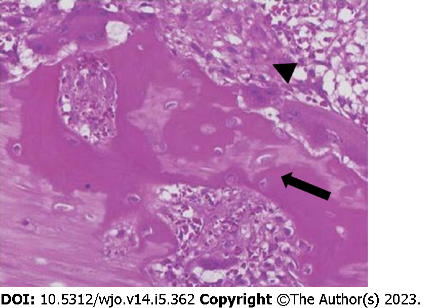
Figure 3 Microscopic findings of myositis ossificans.
Haematoxylin and eosin staining showing a mature lamellar bone neo-formation (arrow) and fibroblasts with elongated nuclei (arrowhead) arranged in short irregular fascicles with the typical "zonation pattern" placed in loose myxoid or collagenous stroma. Occasional mitoses can be observed, in the absence of cellular atypia.
Figure 4 The computed tomography scan.
A: An inhomogeneous pelvic mass measuring almost 20 cm in diameter; B: A mass with multiple haemorrhagic areas (arrows); C: A huge mass filling almost all the abdominal cavity and infiltrating the aortic plane and both ureters.
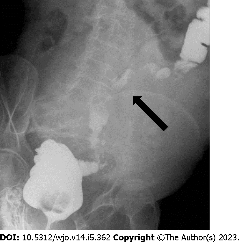
Figure 5 Barium enema showing compression of the sigmoid colon (arrow).
- Citation: Carbone G, Andreasi V, De Nardi P. Intra-abdominal myositis ossificans - a clinically challenging disease: A case report. World J Orthop 2023; 14(5): 362-368
- URL: https://www.wjgnet.com/2218-5836/full/v14/i5/362.htm
- DOI: https://dx.doi.org/10.5312/wjo.v14.i5.362


