Copyright
©The Author(s) 2017.
World J Clin Oncol. Jun 10, 2017; 8(3): 178-189
Published online Jun 10, 2017. doi: 10.5306/wjco.v8.i3.178
Published online Jun 10, 2017. doi: 10.5306/wjco.v8.i3.178
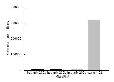
Figure 1 IlluminaHiSeq miRNA expression of tumors tissues biopsies from pancreatic cancer patients.
The data sets used were generated from the the Cancer Genome Atlas Database[71]. The oncogenic miR21 is highly expressed in PC while there is a low expression of tumor suppressor miR200a/b/c (pancreatic cancer samples n = 45).
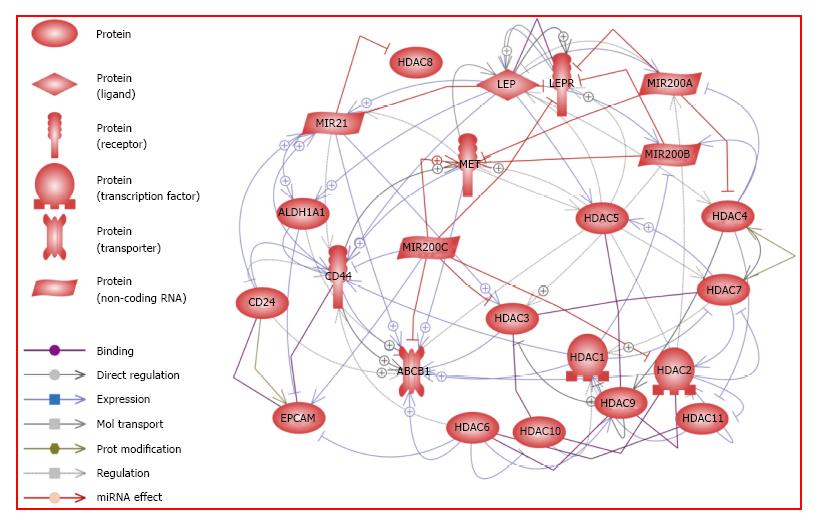
Figure 2 Potential crosstalk between leptin signaling, cancer stem cells, histone deacetylases and microRNA.
Leptin and its receptor LEPR (OB-R) are involved in the regulation of PCSC markers, Classical HDAC, miR21, and miR200a/b/c. HDAC 1, 2, 3, 8: Histone Deacetylases Class I; HDAC 4, 5, 7, 9: Histone Deacetylases Class IIA; HDAC 6,10: Histone Deacetylases Class IIB; HDAC11: Histone Deacetylase Class IV. MiR200a, miR200b, and miR200c: Tumor Suppressors MicroRNA; MiR21: Oncogenic MicroRNA. CD24, CD44, ALDH1A1, ABCB1, MET, and EPCAM: Pancreatic Cancer Stem Cell Markers. HGF: Hepatocyte growth factor cytokine; LEP: Leptin adipokine; LEPR: Leptin receptor. Data generated from Pathway Studio (Pathway Studio – web; Ariadne Genomics, Inc.). Genes were analyzed by Pathway Studio 11 software (Elsevier, Inc., Atlanta, GA, United States) for disease, cellular processes and miRNA interactions. Only genes that had a P value of 0.05 were reported in this study. Specific references supporting these relationships are shown in Supplemental Table 1. References found by Pathway Studio were exported into an Excel file, column D, that contains the PMID number for the citations.
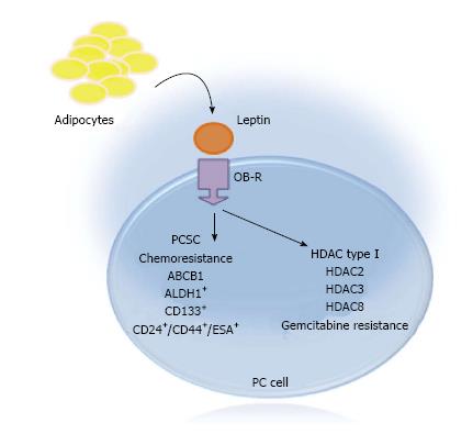
Figure 3 Leptin effects on pancreatic cancer stem cells and histone deacetylases in pancreatic cancer tumorspheres.
Representative cartoon of the effects of leptin on PC cells in vitro. Leptin induced the expression of PCSC markers (CD24+/CD44+/ESA+, CD133+ and ALDH1+). Leptin also increased the levels of ABCB1 [P-glycoprotein 1 or multidrug resistance protein 1 (MDR1) or ATP-binding cassette sub-family B member 1], which is involved in chemoresistance. Additionally, leptin induced the expression of HDAC type I (HDAC 2, 3 and 8). Leptin attenuates the cytotoxic effects of gemcitabine on PC. PC cells were cultured in low attachment plates containing mammocult media (Stem Cell Technol.), which allow the growth of tumorspheres. The tumorspheres were treated for 6 d with leptin (1.2 nmol/L), IONP-LPrA2 (a leptin antagonist bound to iron oxide nanoparticles; 0.0072 pmol/L), and gemcitabine (2 μmol/L). PC viability, PCSC markers and HDAC expression were determined by flow cytometry. Experiments were repeated three times[32,33,81,95].
- Citation: Tchio Mantho CI, Harbuzariu A, Gonzalez-Perez RR. Histone deacetylases, microRNA and leptin crosstalk in pancreatic cancer. World J Clin Oncol 2017; 8(3): 178-189
- URL: https://www.wjgnet.com/2218-4333/full/v8/i3/178.htm
- DOI: https://dx.doi.org/10.5306/wjco.v8.i3.178


