Copyright
©The Author(s) 2016.
World J Gastrointest Pathophysiol. Nov 15, 2016; 7(4): 300-306
Published online Nov 15, 2016. doi: 10.4291/wjgp.v7.i4.300
Published online Nov 15, 2016. doi: 10.4291/wjgp.v7.i4.300
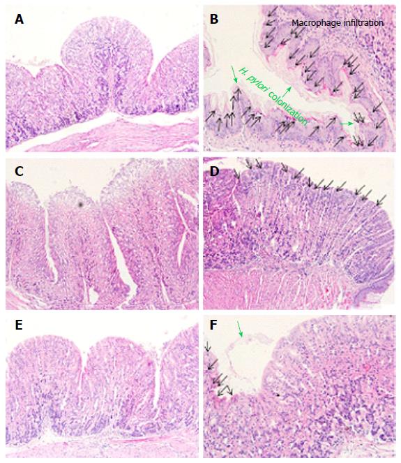
Figure 4 Histology of the gastric mucosa.
Mouse stomachs were removed and underwent hematoxylin and eosin staining. H. pylori colonization and macrophage infiltration were observed under a light microscope. A: Normal group; B: H. pylori infected group; C: Antibiotics treated group; D: S-NANA treated group; E: G-NANA treated group; F: CaG-NANA treated group. S-NANA: Standard N-acetylneuraminic acid; G-NANA: Glycomacropeptide-N-acetylneuraminic acid; CaG-NANA: Calcium-glycomacropeptide-N-acetylneuraminic acid.
- Citation: Rhee YH, Ku HJ, Noh HJ, Cho HH, Kim HK, Ahn JC. Anti-Helicobacter pylori effect of CaG-NANA, a new sialic acid derivative. World J Gastrointest Pathophysiol 2016; 7(4): 300-306
- URL: https://www.wjgnet.com/2150-5330/full/v7/i4/300.htm
- DOI: https://dx.doi.org/10.4291/wjgp.v7.i4.300


