
















Copyright: ©Author(s) 2026.
World J Gastrointest Pathophysiol. Mar 22, 2026; 17(1): 115307
Published online Mar 22, 2026. doi: 10.4291/wjgp.v17.i1.115307
Published online Mar 22, 2026. doi: 10.4291/wjgp.v17.i1.115307
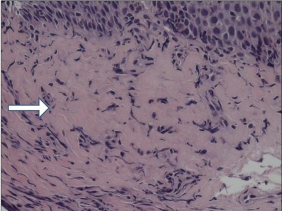
Figure 3 Esophageal epithelium in long-standing eosinophilic esophagitis.
Microphotograph shows a marked increase of fibrotic tissue in the subepithelial layer (white arrow)[22]. Citation: Straumann A, Schoepfer A. Update on basic and clinical aspects of eosinophilic oesophagitis. Gut 2014; 63: 1355-1363. Copyright© The Authors 2026. Published by BMJ Publishing Group. The authors have obtained the permission (Supplementary material).
- Citation: Qasim A, Zacharia GS, Veena F, Kandhi SD, Patel H. From esophagus to colon: A narrative review of eosinophilic gastrointestinal disorders. World J Gastrointest Pathophysiol 2026; 17(1): 115307
- URL: https://www.wjgnet.com/2150-5330/full/v17/i1/115307.htm
- DOI: https://dx.doi.org/10.4291/wjgp.v17.i1.115307