Copyright
©The Author(s) 2017.
World J Radiol. Dec 28, 2017; 9(12): 454-458
Published online Dec 28, 2017. doi: 10.4329/wjr.v9.i12.454
Published online Dec 28, 2017. doi: 10.4329/wjr.v9.i12.454
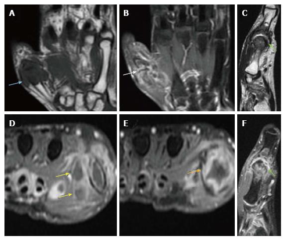
Figure 4 Plain and contrast enhanced magnetic resonance imaging images of proximal hand.
A: Coronal T1W. Hypointense soft-tissue is noted surrounding the first MCP joint with hypointense marrow changes; B: Coronal STIR. Irregular hyperintensities are seen surrounding the first MCP joint along with hyperintensity within the peri-articular bone marrow; C: Sagittal T2W. Mild joint effusion is seen with hypertrophied T2 hypointense synovium and pannus formation; D-F: Axial and sagittal post-contrast Fat Sat T1W. Arrows indicate peripherally enhancing abscesses adjacent to the first MCP joint. Also noted in the vicinity is enhancing proliferative pannus. MCP: Metacarpophalangeal.
- Citation: Mahajan A, Santhoshkumar GV, Kawthalkar AS, Vaish R, Sable N, Arya S, Desai S. Case of victims of modern imaging technology: Increased information noise concealing the diagnosis. World J Radiol 2017; 9(12): 454-458
- URL: https://www.wjgnet.com/1949-8470/full/v9/i12/454.htm
- DOI: https://dx.doi.org/10.4329/wjr.v9.i12.454


