Copyright
©The Author(s) 2017.
World J Radiol. Dec 28, 2017; 9(12): 454-458
Published online Dec 28, 2017. doi: 10.4329/wjr.v9.i12.454
Published online Dec 28, 2017. doi: 10.4329/wjr.v9.i12.454
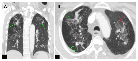
Figure 3 Computed tomography thorax images of the positron emission tomography-computed tomography scan.
A: Coronal CT scan image of the thorax in lung window; B: Axial CT scan image of the thorax in lung window at upper thorax. These images show ground glass opacities (red arrow) with multiple soft tissue density nodules (green arrow), fibrotic changes and calcified granulomas in both the lungs. Features are suggestive of active tuberculosis. CT: Computed tomography.
- Citation: Mahajan A, Santhoshkumar GV, Kawthalkar AS, Vaish R, Sable N, Arya S, Desai S. Case of victims of modern imaging technology: Increased information noise concealing the diagnosis. World J Radiol 2017; 9(12): 454-458
- URL: https://www.wjgnet.com/1949-8470/full/v9/i12/454.htm
- DOI: https://dx.doi.org/10.4329/wjr.v9.i12.454


