Copyright
©The Author(s) 2017.
World J Radiol. Dec 28, 2017; 9(12): 454-458
Published online Dec 28, 2017. doi: 10.4329/wjr.v9.i12.454
Published online Dec 28, 2017. doi: 10.4329/wjr.v9.i12.454
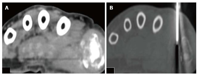
Figure 2 Axial computed tomography of the hand.
The patient is positioned in prone position. A: Axial CT scan image in soft tissue window; B: Axial CT scan image in bone window is showing Trucut biopsy needle taking tissue sample from the soft tissue component surrounding the area of lytic destructive lesion at the first metacarpo-phalangeal joint. CT: Computed tomography.
- Citation: Mahajan A, Santhoshkumar GV, Kawthalkar AS, Vaish R, Sable N, Arya S, Desai S. Case of victims of modern imaging technology: Increased information noise concealing the diagnosis. World J Radiol 2017; 9(12): 454-458
- URL: https://www.wjgnet.com/1949-8470/full/v9/i12/454.htm
- DOI: https://dx.doi.org/10.4329/wjr.v9.i12.454


