Copyright
©The Author(s) 2017.
World J Radiol. Dec 28, 2017; 9(12): 438-447
Published online Dec 28, 2017. doi: 10.4329/wjr.v9.i12.438
Published online Dec 28, 2017. doi: 10.4329/wjr.v9.i12.438
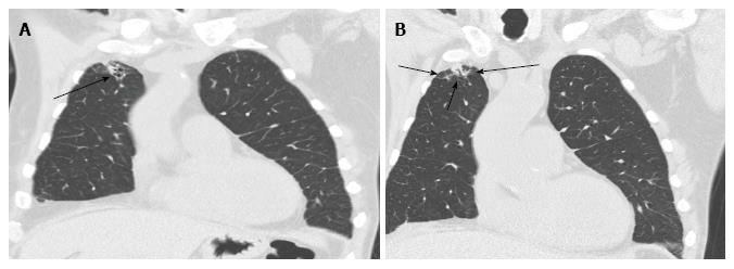
Figure 15 Broncho-alveolar lavage proven aspergillosis.
CT scan of the chest shows right lung apex sub-pleural nodule with surrounding ground glass opacity and focal bronchiectasis. A: Coronal slice of CT chest showing right apical sub-pleural nodule (arrow); B: Coronal slice of CT chest showing surrounding ground glass opacity and focal bronchiectasis (arrow) around a sub-pleural nodule. CT: Computerised tomography.
- Citation: Chia E, Babawale SN. Imaging features of intrathoracic complications of lung transplantation: What the radiologists need to know. World J Radiol 2017; 9(12): 438-447
- URL: https://www.wjgnet.com/1949-8470/full/v9/i12/438.htm
- DOI: https://dx.doi.org/10.4329/wjr.v9.i12.438


