Copyright
©The Author(s) 2017.
World J Radiol. Dec 28, 2017; 9(12): 438-447
Published online Dec 28, 2017. doi: 10.4329/wjr.v9.i12.438
Published online Dec 28, 2017. doi: 10.4329/wjr.v9.i12.438
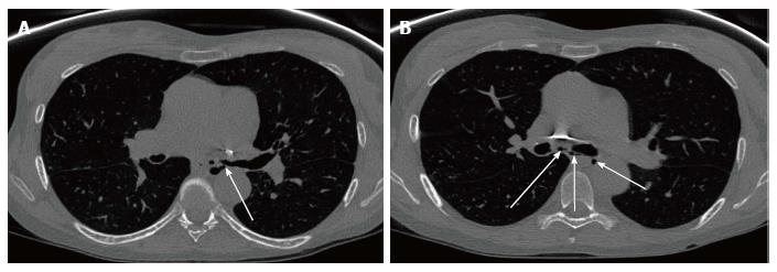
Figure 9 Computerised tomography scan of the chest in a 51-year-old female performed nearly 3 years post bilateral lung transplantation shows left main bronchial dehiscence resulting in gas locules tracking from the left main stem bronchus to the mediastinum causing pneumomediastinum.
A: Axial slice of CT chest image showing gas leaks (arrow) from the left main stem bronchus; B: Axial slice of CT chest image showing multiple gas locules (arrow) causing pneumomediastinum. CT: Computerised tomography.
- Citation: Chia E, Babawale SN. Imaging features of intrathoracic complications of lung transplantation: What the radiologists need to know. World J Radiol 2017; 9(12): 438-447
- URL: https://www.wjgnet.com/1949-8470/full/v9/i12/438.htm
- DOI: https://dx.doi.org/10.4329/wjr.v9.i12.438


