Copyright
©The Author(s) 2016.
World J Radiol. Sep 28, 2016; 8(9): 799-808
Published online Sep 28, 2016. doi: 10.4329/wjr.v8.i9.799
Published online Sep 28, 2016. doi: 10.4329/wjr.v8.i9.799
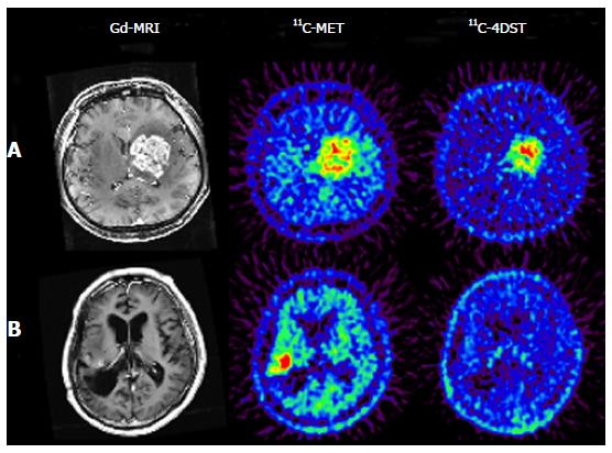
Figure 4 Brain tumor imaging in temozolomide-resistant (A) and -responsive (B) patients.
DNA synthesis images provide completely different information from those obtained with an amino acid transport agent. In the case of recurrent anaplastic oligodendroglioma (A), both 11C-MET and 11C-4DST showed high uptake in the gadolinium-enhanced region of the MRI. However, the distribution pattern of each tracer in the tumor region was not identical. The tumor in this patient showed progressive enlargement despite continuous treatment with the DNA alkylating agent temozolomide. In the case of recurrent anaplastic astrocytoma (B), the patient received one course of treatment with temozolomide 3 d before PET examinations, and it was found that the 11C-4DST uptake was negligible in the gadolinium-enhanced region where high uptake of 11C-MET was observed. The enhanced tumor mass in this patient remained unchanged over 6 mo after commencement of temozolomide treatment. These observations suggest that DNA synthesis in the tumor was suspended by temozolomide treatment. Reprinted from Nariai et al[14], with the permission of Springer. 11C-4DST: 4’-[methyl-11C]-thiothymidine; 11C-MET: 11C-Methionine; Gd-MRI: Gadolinium-enhanced magnetic resonance image; PET: Positron emission tomography.
- Citation: Toyohara J. Evaluation of DNA synthesis with carbon-11-labeled 4′-thiothymidine. World J Radiol 2016; 8(9): 799-808
- URL: https://www.wjgnet.com/1949-8470/full/v8/i9/799.htm
- DOI: https://dx.doi.org/10.4329/wjr.v8.i9.799


