Copyright
©The Author(s) 2016.
World J Radiol. Sep 28, 2016; 8(9): 799-808
Published online Sep 28, 2016. doi: 10.4329/wjr.v8.i9.799
Published online Sep 28, 2016. doi: 10.4329/wjr.v8.i9.799
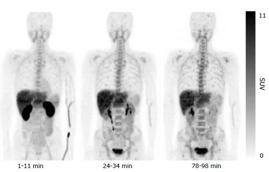
Figure 3 Representative whole-body decay-corrected maximum intensity-projection images of 4’-[methyl-11C]-thiothymidine positron emission tomography images.
11C-4DST showed high uptake in the excretory organs, such as the kidneys, liver, and urinary bladder. Moderate uptake was observed in the proliferative organs, such as bone marrow, spleen, and small intestine. The lowest uptake was observed in the non-proliferating tissues, such as muscle and lungs. This research was originally published in JNM[10]. 11C-4DST: 4’-[methyl-11C]-thiothymidine; SUV: Standardized uptake value.
- Citation: Toyohara J. Evaluation of DNA synthesis with carbon-11-labeled 4′-thiothymidine. World J Radiol 2016; 8(9): 799-808
- URL: https://www.wjgnet.com/1949-8470/full/v8/i9/799.htm
- DOI: https://dx.doi.org/10.4329/wjr.v8.i9.799


