Copyright
©The Author(s) 2016.
World J Radiol. Jul 28, 2016; 8(7): 656-667
Published online Jul 28, 2016. doi: 10.4329/wjr.v8.i7.656
Published online Jul 28, 2016. doi: 10.4329/wjr.v8.i7.656
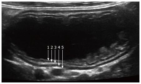
Figure 1 Normal small bowel.
Ultrasound image of small bowel obtained after ingestion of water, using high-resolution linear probe. Five wall layers include: 1-mucosal interface with lumen (hyperechoic), 2-mucosa (hypoechoic), 3-submucosa (hyperechoic), 4-muscularis (hypoechoic), and 5-serosa (hyperechoic).
- Citation: Gale HI, Gee MS, Westra SJ, Nimkin K. Abdominal ultrasonography of the pediatric gastrointestinal tract. World J Radiol 2016; 8(7): 656-667
- URL: https://www.wjgnet.com/1949-8470/full/v8/i7/656.htm
- DOI: https://dx.doi.org/10.4329/wjr.v8.i7.656


