Copyright
©The Author(s) 2016.
World J Radiol. Jun 28, 2016; 8(6): 635-655
Published online Jun 28, 2016. doi: 10.4329/wjr.v8.i6.635
Published online Jun 28, 2016. doi: 10.4329/wjr.v8.i6.635
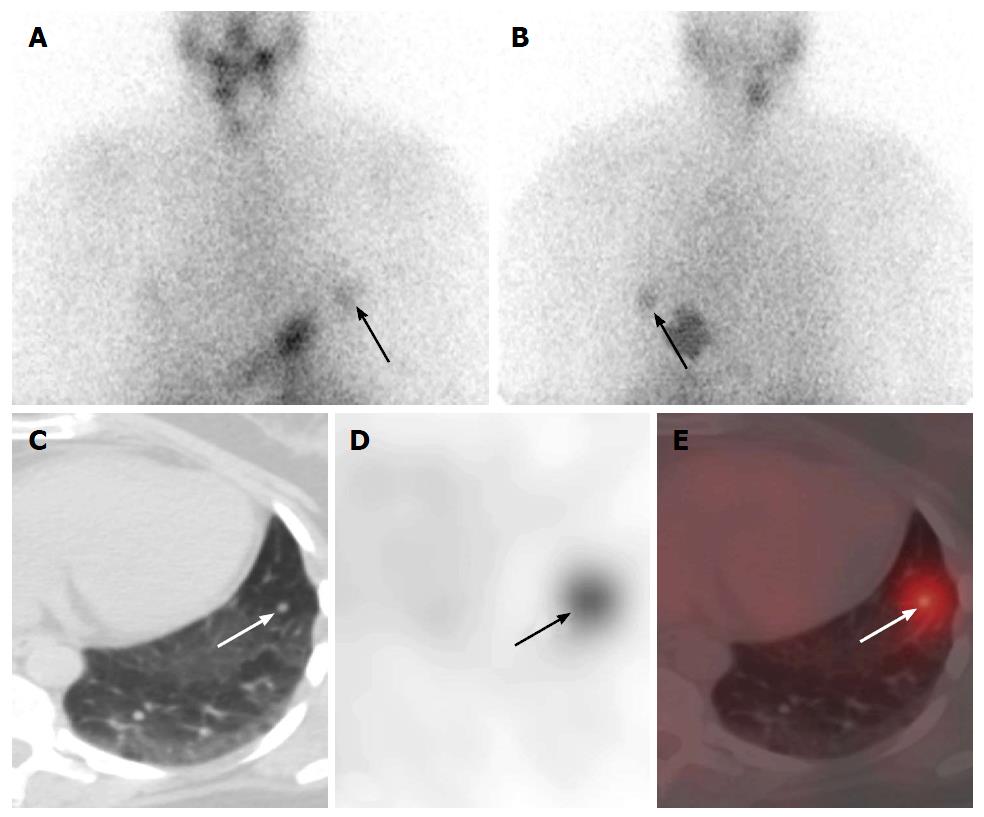
Figure 3 Pre-ablation hypothyroid 131I-radioiodine whole body planar and single photon emission computed tomography-computed tomography scan in a 45-year-old female with papillary thyroid cancer after total thyroidectomy.
Anterior (A) and posterior (B) planar images demonstrate a focus of activity in the left thorax (arrow), which is better visualized on the posterior image. Unenhanced axial CT (C), SPECT (D), and SPECT-CT (E) show a 7 mm a radioiodine avid nodule in the lingula (arrow). The utilization of SPECT-CT increases diagnostic certainty in comparison to planar-only imaging evaluation, obviates the need for concurrent diagnostic CT and allows therapeutic evaluation of this nodule after radioactive iodine thyroid treatment. SPECT: Single photon emission computed tomography; CT: Computed tomography.
- Citation: Wong KK, Gandhi A, Viglianti BL, Fig LM, Rubello D, Gross MD. Endocrine radionuclide scintigraphy with fusion single photon emission computed tomography/computed tomography. World J Radiol 2016; 8(6): 635-655
- URL: https://www.wjgnet.com/1949-8470/full/v8/i6/635.htm
- DOI: https://dx.doi.org/10.4329/wjr.v8.i6.635


