Copyright
©The Author(s) 2016.
World J Radiol. Jun 28, 2016; 8(6): 610-617
Published online Jun 28, 2016. doi: 10.4329/wjr.v8.i6.610
Published online Jun 28, 2016. doi: 10.4329/wjr.v8.i6.610
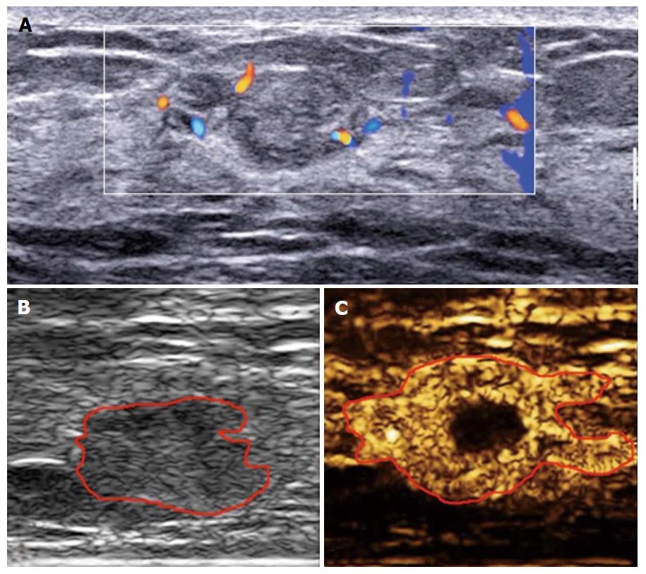
Figure 3 Enhancement patterns of inflammatory lesion.
A: Color Doppler flow imaging with hyper-vascular; B: Heterogeneous hyper-enhancement with rapid wash-in, enlarged size compared with 2 dimensional image, with perfusion defect, irregular shape and unclear margin, without penetrating vessel and crab claw-like pattern.
- Citation: Luo J, Chen JD, Chen Q, Yue LX, Zhou G, Lan C, Li Y, Wu CH, Lu JQ. Contrast-enhanced ultrasound improved performance of breast imaging reporting and data system evaluation of critical breast lesions. World J Radiol 2016; 8(6): 610-617
- URL: https://www.wjgnet.com/1949-8470/full/v8/i6/610.htm
- DOI: https://dx.doi.org/10.4329/wjr.v8.i6.610


