Copyright
©The Author(s) 2016.
World J Radiol. Jun 28, 2016; 8(6): 537-555
Published online Jun 28, 2016. doi: 10.4329/wjr.v8.i6.537
Published online Jun 28, 2016. doi: 10.4329/wjr.v8.i6.537
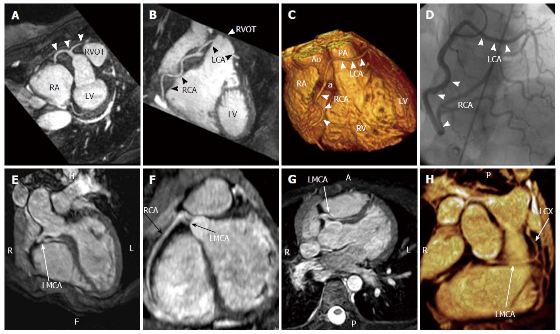
Figure 13 Cases of single coronary artery.
A-D: Coronary cardiac magnetic resonance, T2-prepared navigator-gated whole-heart technique shows the anomalous origin of a single coronary artery from the right sinus of Valsalva in a patient complaining of angina. A: Maximum intensity projection (MIP) image demonstrating the anomalous origin of the single coronary artery from the right sinus of Valsalva (white arrows); B: MIP image showing the path of the right coronary artery and of the left coronary artery, crossing in front of the right ventricle outflow tract; C: Volume rendering reconstruction (VRT) of the heart, showing the anomalous origin of the single coronary artery (a) from the aorta and the course of the RCA and the LCA; D: Coronary angiography in right anterior oblique projection, showing the anomalous coronary tree[17,18]; E-H: Case of single coronary artery arising from the right sinus of Valsalva with interarterial course of the left main coronary artery and distal origin of left circumflex (arrows) seen on three-dimensional cardiac magnetic resonance multiplanar reconstruction, MIP and volume rendering images[19]. Ao: Aorta; LCA: Left coronary artery; PA: Pulmonary artery; RVOT: Right ventricle outflow tract; RA: Right atrium; LMCA: Left main coronary artery; LCX: Left circumflex artery; RCA: Right coronary artery; LV: Left ventricle; RV: Right ventricle.
- Citation: Villa AD, Sammut E, Nair A, Rajani R, Bonamini R, Chiribiri A. Coronary artery anomalies overview: The normal and the abnormal. World J Radiol 2016; 8(6): 537-555
- URL: https://www.wjgnet.com/1949-8470/full/v8/i6/537.htm
- DOI: https://dx.doi.org/10.4329/wjr.v8.i6.537


