Copyright
©The Author(s) 2016.
World J Radiol. Mar 28, 2016; 8(3): 298-307
Published online Mar 28, 2016. doi: 10.4329/wjr.v8.i3.298
Published online Mar 28, 2016. doi: 10.4329/wjr.v8.i3.298
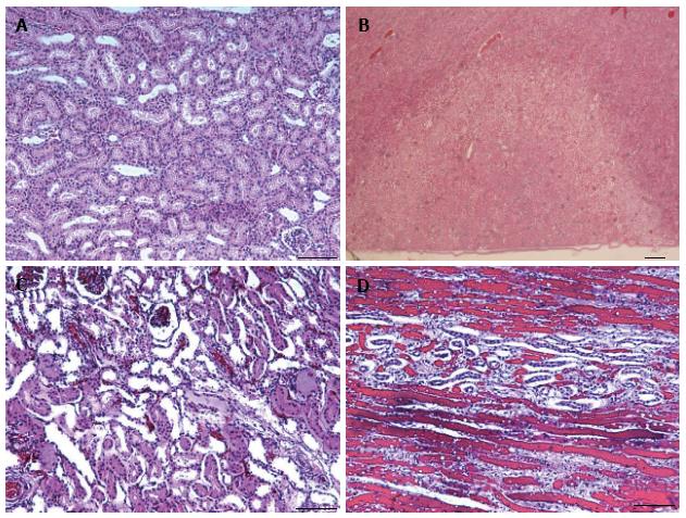
Figure 8 Microscopic sections (hematoxylin-eosin) of a normal renal tissue (A, × 40), the territory of both ablated (pale) and normal renal tissue (B, × 10) and ablated lesion (C, × 40) with damaged nephrons in a haphazard array and hemorrhage (D, × 100).
Note the close agreement in the triangular infarct territory on microscopy and microscopy lesions in Figure 6 and magnetic resonance imaging in Figure 6. There was no evidence of peri-renal tissue injury (B).
- Citation: Saeed M, Krug R, Do L, Hetts SW, Wilson MW. Renal ablation using magnetic resonance-guided high intensity focused ultrasound: Magnetic resonance imaging and histopathology assessment. World J Radiol 2016; 8(3): 298-307
- URL: https://www.wjgnet.com/1949-8470/full/v8/i3/298.htm
- DOI: https://dx.doi.org/10.4329/wjr.v8.i3.298


