Copyright
©The Author(s) 2016.
World J Radiol. Feb 28, 2016; 8(2): 183-191
Published online Feb 28, 2016. doi: 10.4329/wjr.v8.i2.183
Published online Feb 28, 2016. doi: 10.4329/wjr.v8.i2.183
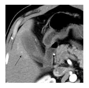
Figure 8 Axial arterial phase computed tomography image showing an area of hyper perfusion in the segment V of liver adjoining a gallbladder showing diffusely thickened walls.
Also noted is the blurring of interfaces between gallbladder wall and adjoining liver parenchyma. Liver infiltration can demonstrate an early enhancement of the parenchyma which pathologically corresponds with accumulation of inflammatory cells and abundant fibrosis.
- Citation: Singh VP, Rajesh S, Bihari C, Desai SN, Pargewar SS, Arora A. Xanthogranulomatous cholecystitis: What every radiologist should know. World J Radiol 2016; 8(2): 183-191
- URL: https://www.wjgnet.com/1949-8470/full/v8/i2/183.htm
- DOI: https://dx.doi.org/10.4329/wjr.v8.i2.183


