Copyright
©The Author(s) 2016.
World J Radiol. Feb 28, 2016; 8(2): 183-191
Published online Feb 28, 2016. doi: 10.4329/wjr.v8.i2.183
Published online Feb 28, 2016. doi: 10.4329/wjr.v8.i2.183
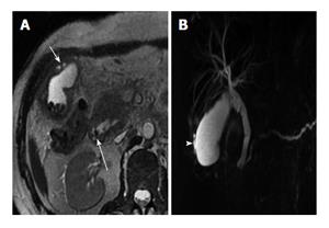
Figure 7 Axial T2W image (A) and corresponding magnetic resonance cholangiopancreatography image (B).
Axial T2W image (A) showing multiple gallbladder calculi with a diffusely thickened wall showing multiple intramural nodules (short arrow). Note is also made of a small filling defect involving the ampullary common duct (long arrow). Corresponding MRCP image (B) showing multiple intramural nodules (white arrowhead) along with a dilated pancreatico-biliary system secondary to calculus in ampullary region. MRCP: Magnetic resonance cholangiopancreatography.
- Citation: Singh VP, Rajesh S, Bihari C, Desai SN, Pargewar SS, Arora A. Xanthogranulomatous cholecystitis: What every radiologist should know. World J Radiol 2016; 8(2): 183-191
- URL: https://www.wjgnet.com/1949-8470/full/v8/i2/183.htm
- DOI: https://dx.doi.org/10.4329/wjr.v8.i2.183


