Copyright
©The Author(s) 2016.
World J Radiol. Feb 28, 2016; 8(2): 183-191
Published online Feb 28, 2016. doi: 10.4329/wjr.v8.i2.183
Published online Feb 28, 2016. doi: 10.4329/wjr.v8.i2.183
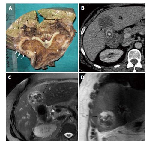
Figure 3 Rupture of gallbladder serosal lining and spread of the inflammatory response leads to adhesions with adjacent liver, duodenum and transverse colon.
Gross pathology specimen (A) demonstrating a thickened gallbladder wall (dotted arrow) showing multiple yellowish nodules (xanthoma nodules) within (short arrows). Infiltration into the adjoining liver parenchyma is seen (black arrows). Black arrowheads denote the normal liver parechyma while the white arrowheads denote gallbladder lumen. Corresponding computed tomography images (B) of the same patient showing focal mural thickening involving the gallbladder fundal region with poor fat planes and infiltration into the adjacent hepatic parenchyma (black arrows). Magnetic resonance axial and coronal images (C and D) of the same patient showing heterogeneous mass like thickening involving the gallbladder fundal region with poor fat planes to adjacent hepatic parenchyma (black arrows).
- Citation: Singh VP, Rajesh S, Bihari C, Desai SN, Pargewar SS, Arora A. Xanthogranulomatous cholecystitis: What every radiologist should know. World J Radiol 2016; 8(2): 183-191
- URL: https://www.wjgnet.com/1949-8470/full/v8/i2/183.htm
- DOI: https://dx.doi.org/10.4329/wjr.v8.i2.183


