Copyright
©The Author(s) 2015.
World J Radiol. Aug 28, 2015; 7(8): 212-219
Published online Aug 28, 2015. doi: 10.4329/wjr.v7.i8.212
Published online Aug 28, 2015. doi: 10.4329/wjr.v7.i8.212
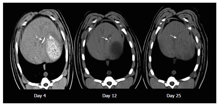
Figure 5 Serial non-enhanced computed tomography scans of pig’s liver taken on 4, 12, and 25 d after the microsphere embolization showing its radiopaque characteristic and the gradual fade along with time (white arrow).
- Citation: Liu YS, Lin XZ, Tsai HM, Tsai HW, Chen GC, Chen SF, Kang JW, Chou CM, Chen CY. Development of biodegradable radiopaque microsphere for arterial embolization-a pig study. World J Radiol 2015; 7(8): 212-219
- URL: https://www.wjgnet.com/1949-8470/full/v7/i8/212.htm
- DOI: https://dx.doi.org/10.4329/wjr.v7.i8.212


