Copyright
©2014 Baishideng Publishing Group Inc.
World J Radiol. Jul 28, 2014; 6(7): 446-458
Published online Jul 28, 2014. doi: 10.4329/wjr.v6.i7.446
Published online Jul 28, 2014. doi: 10.4329/wjr.v6.i7.446
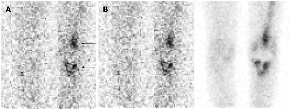
Figure 8 Aseptically loosened left knee arthroplasty.
A: On this anterior image from an indium-111 labeled leukocyte study, there is intense periprosthetic activity around both the tibial and femoral components (arrows), while there is no activity around the contralateral knee. The study could be interpreted erroneously as positive for infection. Compare the intensity of activity around this prosthesis with the intensity of activity around the infected hip arthroplasty in Figure 7A. As these two cases illustrate, the intensity of labeled leukocyte activity around a prosthetic joint is not a reliable criterion for determining the presence or absence of infection; B: The distribution of periprosthetic activity on the labeled leukocyte (left, 111In-WBC) and sulfur colloid bone marrow (right, 99mTc-SC) images is virtually identical (spatially congruent) and the combined study is negative for infection. The periprosthetic activity on the labeled leukocyte image is due to marrow, not to infection. Performing complementary bone marrow imaging eliminates the two major difficulties inherent in the interpretation of labeled leukocyte images: variable intensity of periprosthetic activity and differentiating bone marrow activity from infection. Same patient illustrated in Figure 8A.
- Citation: Palestro CJ. Nuclear medicine and the failed joint replacement: Past, present, and future. World J Radiol 2014; 6(7): 446-458
- URL: https://www.wjgnet.com/1949-8470/full/v6/i7/446.htm
- DOI: https://dx.doi.org/10.4329/wjr.v6.i7.446


