
















©2014 Baishideng Publishing Group Inc.
World J Radiol. Jun 28, 2014; 6(6): 329-343
Published online Jun 28, 2014. doi: 10.4329/wjr.v6.i6.329
Published online Jun 28, 2014. doi: 10.4329/wjr.v6.i6.329
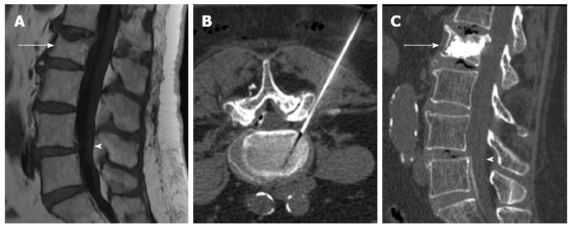
Figure 7 Sagittal T1 weighted image (A) showing vertebral compression fracture of L2 (arrow) and degenerative spondylolisthesis of L4 (arrowhead), computed tomography guided transforaminal epidural injection (B), Sagittal computed tomography after vertebroplasty (arrow) and epidural injection (arrowhead) (C).
- Citation: Santiago FR, Chinchilla AS, Álvarez LG, Abela ALP, García MDMC, López MP. Comparative review of vertebroplasty and kyphoplasty. World J Radiol 2014; 6(6): 329-343
- URL: https://www.wjgnet.com/1949-8470/full/v6/i6/329.htm
- DOI: https://dx.doi.org/10.4329/wjr.v6.i6.329