Copyright
©2014 Baishideng Publishing Group Inc.
World J Radiol. May 28, 2014; 6(5): 203-209
Published online May 28, 2014. doi: 10.4329/wjr.v6.i5.203
Published online May 28, 2014. doi: 10.4329/wjr.v6.i5.203
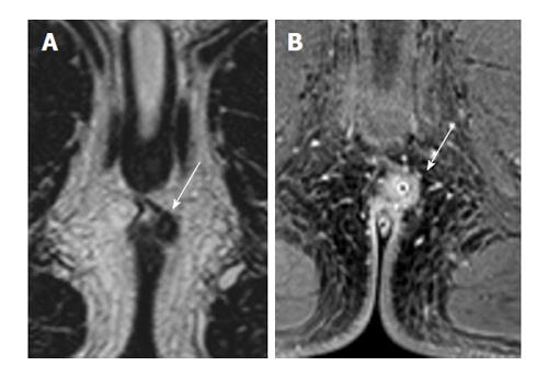
Figure 5 A 20-year-old with intersphincteric fistula (white arrows) treated with seton image.
A: 2D T2 turbo spin-echo; B: 3D T1 weighted prepared gradient echo sequence with fat saturation. The seton is seen clearly on THRIVE, as well as the inflammation, but not on T2. However, the intersphincteric course of the fistula is clearer on T2, while the extent of inflammation obscures the thin external sphincter.
- Citation: Torkzad MR, Ahlström H, Karlbom U. Comparison of different magnetic resonance imaging sequences for assessment of fistula-in-ano. World J Radiol 2014; 6(5): 203-209
- URL: https://www.wjgnet.com/1949-8470/full/v6/i5/203.htm
- DOI: https://dx.doi.org/10.4329/wjr.v6.i5.203


