Copyright
©2014 Baishideng Publishing Group Inc.
World J Radiol. May 28, 2014; 6(5): 203-209
Published online May 28, 2014. doi: 10.4329/wjr.v6.i5.203
Published online May 28, 2014. doi: 10.4329/wjr.v6.i5.203
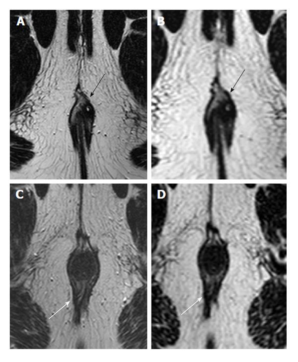
Figure 1 Two different patients imaged with both 2D (A and C) and 3D T2 turbo spin-echo (B and D) images.
In most patients, such as a 30-year-old male with intersphincteric fistula (black arrows), depictions were clear on both 2D (A) and 3D (B). However, in the 39-year-old male depicted in images C and D, the fistula (white arrows) entering dorsally was noticed on 2D images (C), but not those that were 3D (D). Note that there is a seton partially visible inside the tract on the 2D image.
- Citation: Torkzad MR, Ahlström H, Karlbom U. Comparison of different magnetic resonance imaging sequences for assessment of fistula-in-ano. World J Radiol 2014; 6(5): 203-209
- URL: https://www.wjgnet.com/1949-8470/full/v6/i5/203.htm
- DOI: https://dx.doi.org/10.4329/wjr.v6.i5.203


