Copyright
©2014 Baishideng Publishing Group Inc.
World J Radiol. Oct 28, 2014; 6(10): 846-849
Published online Oct 28, 2014. doi: 10.4329/wjr.v6.i10.846
Published online Oct 28, 2014. doi: 10.4329/wjr.v6.i10.846
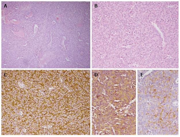
Figure 2 Photomicrograph showing tumor cells arranged in sheets with interspersed blood vessels and areas of necrosis (H and E × 100, A) with small to medium sized cells having round nuclei with stipple chromatin and moderate cytoplasm (H and E × 200, B).
Diffuse membranous immunopositivity for MIC 2 (IHC × 200, C), cytoplasmic immunopositivity for neuron specific enolase (NSE) (IHC × 200, D) and cytoplasmic immunopositivity for Synaptophysin (IHC × 200, E) were also seen. IHC: Immunohistochemistry.
- Citation: Chinnaa S, Das CJ, Sharma S, Singh P, Seth A, Purkait S, Mathur SR. Peripheral primitive neuroectodermal tumor of the kidney presenting with pulmonary tumor embolism: A case report. World J Radiol 2014; 6(10): 846-849
- URL: https://www.wjgnet.com/1949-8470/full/v6/i10/846.htm
- DOI: https://dx.doi.org/10.4329/wjr.v6.i10.846


