
















©2013 Baishideng Publishing Group Co.
World J Radiol. Aug 28, 2013; 5(8): 285-294
Published online Aug 28, 2013. doi: 10.4329/wjr.v5.i8.285
Published online Aug 28, 2013. doi: 10.4329/wjr.v5.i8.285
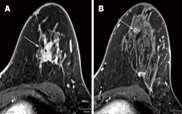
Figure 2 1.
5Tesla axial T1-weighted fat-suppressed contrast enhanced subtraction images in 53-year-old woman with newly diagnosed left breast infiltrating ductal carcinoma show a conglomerate of masses with signal void due to biopsy marker (arrow) in the left superior breast (A), and a mammographically occult rim-enhancing lesion (arrow) 2 cm inferior to the index cancer confirmed as an additional infiltrating ductal carcinoma (B). Note the lower contrast and spatial resolution evident on these images compared to the 3.0Tesla system.
- Citation: Butler RS, Chen C, Vashi R, Hooley RJ, Philpotts LE. 3.0 Tesla vs 1.5 Tesla breast magnetic resonance imaging in newly diagnosed breast cancer patients. World J Radiol 2013; 5(8): 285-294
- URL: https://www.wjgnet.com/1949-8470/full/v5/i8/285.htm
- DOI: https://dx.doi.org/10.4329/wjr.v5.i8.285