
















©2013 Baishideng Publishing Group Co.
World J Radiol. May 28, 2013; 5(5): 215-219
Published online May 28, 2013. doi: 10.4329/wjr.v5.i5.215
Published online May 28, 2013. doi: 10.4329/wjr.v5.i5.215
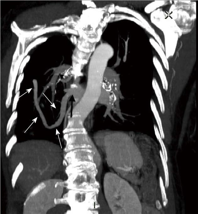
Figure 2 Contrast-enhanced maximum intensity projection multiplanar reformatted coronal image of the computed tomography chest demonstrating the path of the anomalous pulmonary vein (white arrows) and its connection to the left atrium (black arrow).
R: Right.
- Citation: Rodrigues MA, Ritchie G, Murchison JT. Incidental meandering right pulmonary vein, literature review and proposed nomenclature revision. World J Radiol 2013; 5(5): 215-219
- URL: https://www.wjgnet.com/1949-8470/full/v5/i5/215.htm
- DOI: https://dx.doi.org/10.4329/wjr.v5.i5.215