
















©2013 Baishideng Publishing Group Co.
World J Radiol. May 28, 2013; 5(5): 193-201
Published online May 28, 2013. doi: 10.4329/wjr.v5.i5.193
Published online May 28, 2013. doi: 10.4329/wjr.v5.i5.193
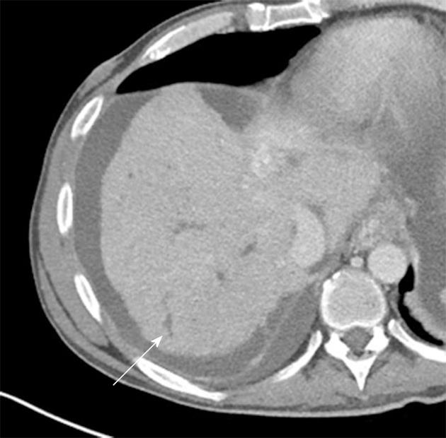
Figure 8 Primary sclerosing cholangitis and Crohn’s disease.
Twenty-four-year-old female with inflammatory bowel disease and primary sclerosing cholangitis. Axial contrast enhanced image demonstrates beading and irregular dilatation of the peripheral intrahepatic biliary tree (arrow). Massive enlargement of the caudate lobe with nodularity of the liver capsule was also noted (not shown).
- Citation: Raman SP, Horton KM, Fishman EK. Computed tomography of Crohn’s disease: The role of three dimensional technique. World J Radiol 2013; 5(5): 193-201
- URL: https://www.wjgnet.com/1949-8470/full/v5/i5/193.htm
- DOI: https://dx.doi.org/10.4329/wjr.v5.i5.193