Copyright
©2013 Baishideng Publishing Group Co.
World J Radiol. Dec 28, 2013; 5(12): 491-497
Published online Dec 28, 2013. doi: 10.4329/wjr.v5.i12.491
Published online Dec 28, 2013. doi: 10.4329/wjr.v5.i12.491
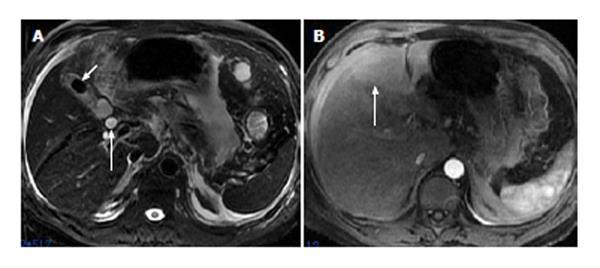
Figure 3 A 49-year-old man with acute pancreatitis combined with cholecystitis, gallbladder stone and common bile duct dilatation.
Respiratory-triggered axial fast recovery fast spin-echo T2-weighted image (A) shows cholecystitis and gallbladder stones (short arrow) as well as common bile duct dilatation (long arrow). Patch- or wedge-shaped hepatic abnormal perfusion is found in the left lobe of the liver on arterial phase images (B, arrow).
- Citation: Tang W, Zhang XM, Zhai ZH, Zeng NL. Hepatic abnormal perfusion visible by magnetic resonance imaging in acute pancreatitis. World J Radiol 2013; 5(12): 491-497
- URL: https://www.wjgnet.com/1949-8470/full/v5/i12/491.htm
- DOI: https://dx.doi.org/10.4329/wjr.v5.i12.491


