
















©2013 Baishideng Publishing Group Co.
World J Radiol. Nov 28, 2013; 5(11): 446-449
Published online Nov 28, 2013. doi: 10.4329/wjr.v5.i11.446
Published online Nov 28, 2013. doi: 10.4329/wjr.v5.i11.446
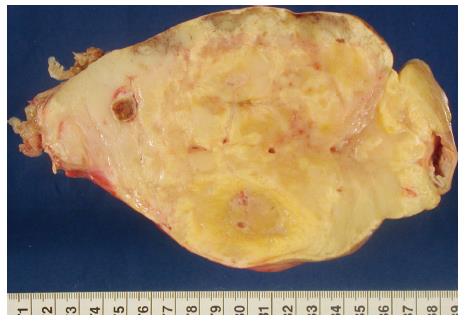
Figure 3 The cut surface of the encapsulated tumor shows a dirty yellow and grey-white color with focal areas of necrosis.
The resected small bowel segment is seen at the edge of the tumor on the right side with no evidence of infiltration.
- Citation: Maataoui A, Khan FM, Vogl TJ, Erler A. Mesenteric myolipoma. World J Radiol 2013; 5(11): 446-449
- URL: https://www.wjgnet.com/1949-8470/full/v5/i11/446.htm
- DOI: https://dx.doi.org/10.4329/wjr.v5.i11.446