
















©2013 Baishideng Publishing Group Co.
World J Radiol. Nov 28, 2013; 5(11): 446-449
Published online Nov 28, 2013. doi: 10.4329/wjr.v5.i11.446
Published online Nov 28, 2013. doi: 10.4329/wjr.v5.i11.446
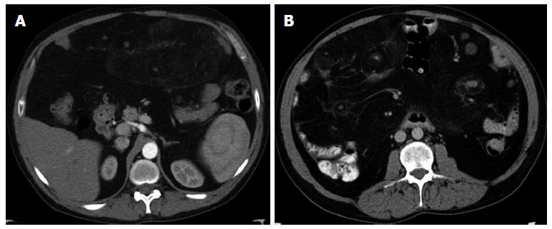
Figure 1 Multidetector computed tomography axial image.
A: The abdomen shows a mesenteric mass measuring 10 cm × 19 cm in its axial orientation with intact capsule (arrowed). The mass is mainly composed of adipose tissue with nodular lesions of soft tissue attenuation; B: The abdomen shows the diffuse nature of the lesion involving the entire mesentery with displacement of the bowel loops to the abdominal wall and separation of the mesenteric vessels.
- Citation: Maataoui A, Khan FM, Vogl TJ, Erler A. Mesenteric myolipoma. World J Radiol 2013; 5(11): 446-449
- URL: https://www.wjgnet.com/1949-8470/full/v5/i11/446.htm
- DOI: https://dx.doi.org/10.4329/wjr.v5.i11.446